Figure 1.21 Helical structure in a CLC microdroplet. Image of microdroplet in a lasing condition is also shown [76].
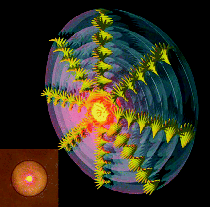
Figure 2.20 Top: Synchrotron XRD patterns from homeotropic monodomain of material 35 (a) and the blend of 35 (b) with PC61BM in an 8-µm-thick glass cell. Bottom: Calculated geometric dimensions of porphyrin 35 and 3D ChemDraw spacing-filling model of fullerene derivative PC61BM and the schematic representations of homeotropically aligned architecture of the blend of 35 and PC61BM. Reproduced with permission from ref. 110.
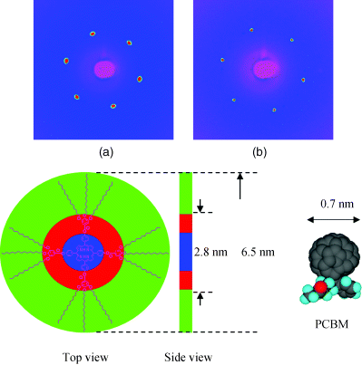
Figure 2.32 Formation of homeotropic texture on a macroscopic scale via slow cooling of the 1.8 µm cell with 33. Slow cooling induces selective nucleation and growth of homeotropic domain (parts (a) and (b) at 121.0°C) and hence yields uniform homeotropic columnar orientation at lower temperatures ((c) 113.0°C and (d) 75.0°C), confirmed by both (e) optical texture under crossed polarizers and (f) conoscopic image at 75.0°C. Optical images were taken at (a–d) 70° and (e) 90° angles between polarizers. The scale bar corresponds to 50 µm. (g) X-ray 2D pattern for the Colh phase at 75.0°C of 33. (h) The azimuthal scan of the peak in (g) shows equally spaced six peaks with uniform intensity distribution. Reproduced with permission from ref. 77(a).
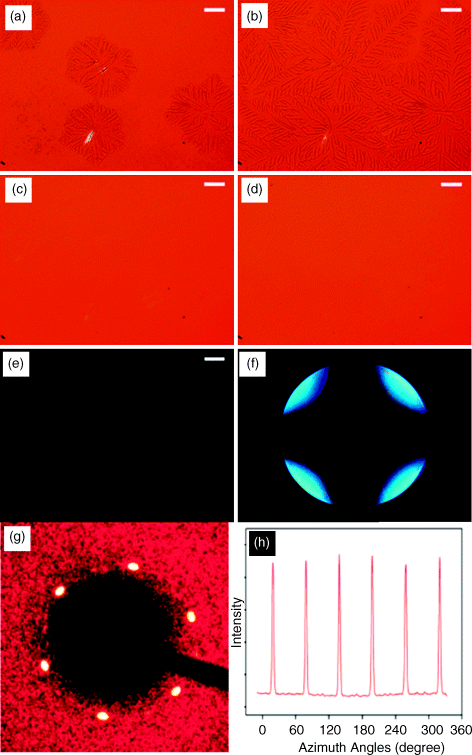
Figure 4.8 Schematic illustration of (a) homeotropically aligned FLC cell with the spontaneous polarization P parallel to the polar axis which is the Y-axis in the XYZ coordinate system and the y-axis in the xyz molecular coordinate system, (b) SHG experiments using phase matching method, and (c) SHG experiments using Maker fringe method at normal incidence. In (b), the phase matching is achieved by rotating the cell around the polar axis, θ is the tilt angle, σi is the incident angle, and σ is the angle between the optical propagation direction and the FLC molecule director ![]() .
.
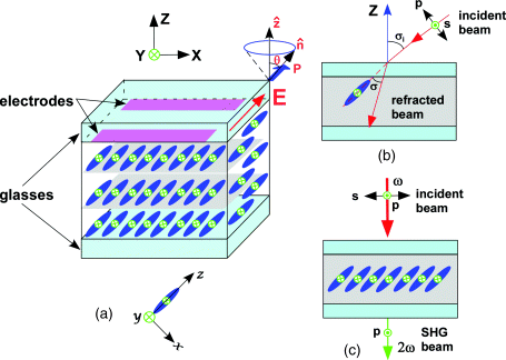
Figure 4.15 Switching by molecular rotation around the long axis (left) changes both chirality and polarity, while switching on the tilt cone (more common, right) only changes polarity with retention of chirality.
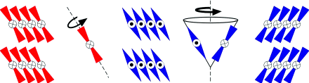
Figure 5.30 (a) Demonstration of photocontraction of a cross-linked polymer liquid crystal containing azobenzene, in which the bending direction of the film is manipulated by the orientation of linearly polarized light in the UV region (inducing contraction) and visible light (recovery of the original shape). Reprinted with permission from ref. 100, Copyright 2003, Nature publishing group. (b) Schematic to illustrate the proposed mechanism governing the photocontraction. Reprinted with permission from ref. 90, Copyright 2006, John Wiley & Sons.
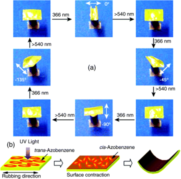
Figure 6.1 A schematic mechanism of the reflective wavelength of light-driven chiral molecular switch or motors in achiral nematic LC media reversibly and dynamically tuned by light.
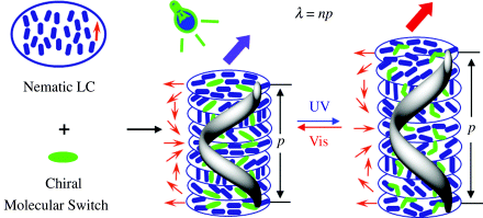
Figure 6.9 Changes in the reflection color of the CLC consisting of chiral azobenzene 9 and non-photoresponsive chiral dopant 10 in E44 by varying UV irradiation time: 0 s (left), 4 s (middle), and 10 s (right) (top); (a) gray mask and (b) red–green–blue (RGB) patterning of the CLC obtained by UV irradiation for 10 s through the gray mask at 25°C. Used with permission from Ref. [43].
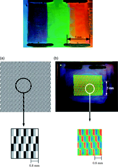
Figure 6.11 A flexible optically addressed photochiral display (A); a conventional display attached bulky and costly electronics compared with an optically addressed display with the same image without the added electronics (B). Used with permission from Ref. [47].
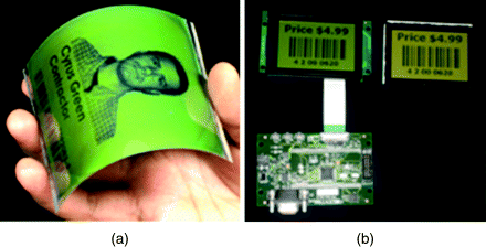
Figure 6.14 Reflection color images of 6.5 wt% chiral switch 2 in commercially available achiral LC host E7 in 5 μm thick planar cell. A: upon UV light at 365 nm (5.0 mW/cm2) with different time; B: reversible back across the entire visible spectrum upon visible light at 520 nm (1.5 mW/cm2) with different time. The colors were taken from a polarized reflective mode microscope. Reflective spectra of 6.5 wt% chiral switch 2 in LC E7 in a 5 μm thick planar cell at room temperature. C: under UV light at 365 nm wavelength (5.0 mW/cm2) with different time (3 s, 8 s, 16 s, 25 s, 40 s, and 47 s, from left to right). D: under visible light at 520 nm wavelength (1.5 mW/cm2) with different time (2 s, 5 s, 9 s, 12 s, and 20 s, from right to left). Used with permission from Ref. [39].
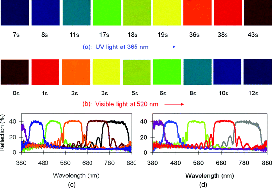
Figure 6.15 mages of 5 μm thick homeotropic alignment cell with 4 wt% chiral switch 2 in LC host E7. (See text for full caption.)
Figure 6.16 Top: Molecular structures of chiral cyclic azobenzenes (R)-17 and (R)-18 (A). Middle (B–D): Schematic mechanism of reflection wavelength tuning and handedness inversion of light-driven chiral molecular switch or motor in achiral nematic LC media reversibly and dynamically tuned by light. Bottom: Polarized optical photomicrographs of a planar aligned N* film containing 10 wt% (R)-17 in ZLI-1132 at room temperature, showing reversible phase transitions occurring by light irradiation of the sample inside a 5 μm cell: (a) oily streak texture of the N* phase before irradiation; (b) N phase obtained by exposure of the sample to UV irradiation; (c) extinguishing orientation of the N cell by rotation between crossed polarizers; (d) regeneration of the oily streak texture of the N* phase upon continued irradiation (bottom–right). Used with permission from Ref. [56]
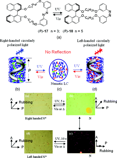
Figure 6.27 Features of a light-driven molecular motor: (a) Molecular structure of chiral motor 47. (b) Polygonal texture of a LC film doped with 1 wt% chiral motor 47. (c) Glass rod rotating on the LC during irradiation with ultraviolet light. (See text for full caption..)
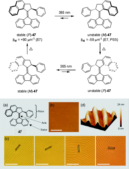
Figure 7.13 The confinement of the cholesteric liquid crystal mixture 1 inside thin cylindrical fibers forces the director helix to expand or compress from its natural pitch, leading to a reflection color Λ that depends on the inner fiber diameter d, as illustrated in the center diagram (black curve). (See text for full caption..)
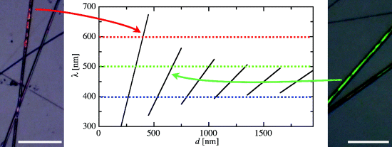
Figure 8.1 (a) Chemical structure and phase transition temperatures of the LC diblock copolymer. (See text for full caption..)
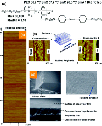
Figure 8.4 (a) Chemical structure and phase transition temperatures of the LC diblock copolymer. (See text for full caption..)
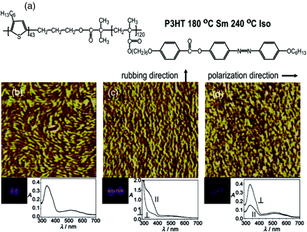
Figure 9.16 Photolithographic process to produce a red, green, and blue pixellated OLED. After patterned irradiation with ultraviolet light, the sample is washed with the solvent used for deposition, so that the unexposed regions are removed.
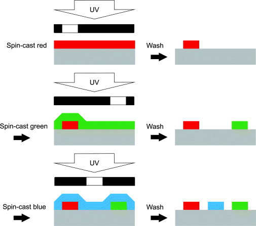
Figure 9.18 A prototype OLED with a red, green, and blue pixel on the same substrate fabricated by sequential spin-coating and polymerization of the materials?20, 5 blend,5 and19 onto a PEDOT:PSS film covering a patterned indium tin oxide (ITO) substrate. (See text for full caption..)
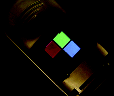
Figure 13.4 Two dimensional X-ray diffraction pattern from (a) isotropic and (b) nematic phases of 4-cyano-4′-pentylbiphenyl (5CB) and (c) cybotactic smectic C phase of nematic phase of bis-(4′-n-octyloxybenzal)-2-chlor1o,4-phenylenediamine. (See text for full caption.)
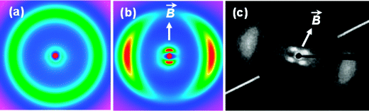
Figure 14.19 Schematic diagram illustrating the formation of vertically aligned graphene layer arrays (a). HR-TEM image showing the full fringe field in Z-axis projection, indicating vertical grapheme layer orientation (b). (See text for full caption.)
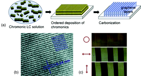
Figure 14.20 The scheme of the LCLC biosensor for the detection of immune complexs. (Redrawn from Shiyanovskii et al. [37].)
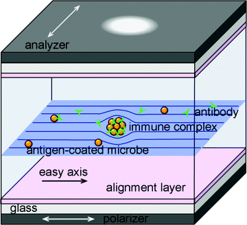
Figure 15.11 (A) and (B) Interference color of a film of nematic 5CB supported on a SAM formed from HOOC(CH2)10SH before (A) and after (B) exposure to n-H2N(CH2)5CH3. (C) Schematic illustration of the orientation of the LC in contact with a carboxylic acid monolayer that is consistent with interference colors shown in panel (A). The bold arrow indicates the direction of deposition of gold onto the substrate. (D) Schematic illustration of the orientation of the LC in contact with the hexylamine-reacted carboxylic acid monolayer that is consistent with the interference colors shown in panel (B). Reprinted with permission from Shah and Abbott [28]). Copyright 2003 American Chemical Society.
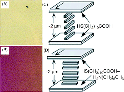
Figure 16.3 Photographs of green house with liquid crystal switchable window in Cleveland Botanic Garden. Photo courtesy of Cleveland Botanic Garden.
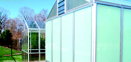
Figure 17.37 The 3D vision sensor camera stage developed.
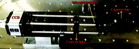
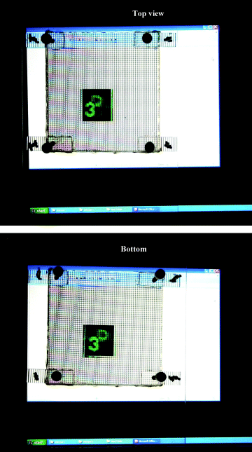
Figure 17.39 The reconstructed 3D image in the developed 3D display from all the elemental images viewed from top and bottom.