
Laser processing of optical fibers: new photosensitivity findings, refractive index engineering and surface structuring
S. Pissadakis, Foundation for Research and Technology – Hellas (FORTH), Institute of Electronic Structure and Laser (IESL), Greece
Abstract:
The chapter presents a selective review on the laser processing of optical fibers, including new photosensitivity findings, refractive index engineering and surface structuring results, reported approximately during the last decade. Topics covered include the Bragg grating fabrication in standard germanosilicate and all-silica glass optical fibers using ultraviolet and infrared lasers with pulse durations from nanosecond to femtosecond, regenerated gratings and inscriptions into ‘soft’ glass fibers. The Bragg grating formation into photonic crystal and microstructured optical fibers is also discussed, while the last part of the chapter focuses onto the end-face, cladding and capillary surface structuring of optical fibers using lasers.
12.1 Introduction and historical overview
The manifold and complex material problem of photosensitivity referring to the underlying physical mechanisms and induced modifications of the physical properties of glasses/optical materials using electromagnetic or hard-particles radiation has remained an active research topic for more than five decades, exhibiting significant engineering impact (Primak and Kampwirth, 1968; Weeks, 1956, 1994). Focusing on glasses, numerous studies have been presented covering a different range of compositions, dopands and exposure conditions/approaches, illustrating the physical processes that take place during irradiation, as well as the corresponding optical, microscopic and mechanical properties modifications induced in the matrices. The favored material which has attracted intense and continuous academic interest is silicate glass, due to its high transparency over a broad band of wavelengths, its mechanical and chemical properties and radiation resistance. Moreover, silicate glasses are the backbone optical materials in numerous everyday applications, in particular those in optical fiber communications and sensing. However, there have also been other matrixes such as those of phosphate, chalcogenide and fluoride glasses that have been studied with respect to their photosensitivity behavior and refractive index changes inscribed.
The invention of laser (Maiman, 1960) and the subsequent development of high power and photon energy laser sources revolutionized the field of optical glasses photosensitivity, efficiently substituting the hard radiation sources used in the decades of the 1950s and 1960s. High power and photon energy laser sources catalyzed light–matter interaction experiments, transferring photosensitivity experiments from the large installation radiation facilities into small-sized laboratories, while increasing engineering investigations and commercialization possibilities. Generally, a laser beam can be easily focused over submicron areas on the surface or in the volume of a glass, interfered, scanned, polarized and scattered, thus becoming an efficient, versatile and potentially low-cost tool for inducing and simultaneously studying photosensitive effects in optical materials. The above capabilities of laser radiation have prompted the evolution of photosensitivity processes in the engineering of refractive index of optical glassy materials over the last 25 years.
Early photosensitivity experiments using hard radiation such as X-rays were performed in silicate bulk glass samples, mostly studying macroscopic modifications induced related to compaction or optical absorption by means of coloration changes. However, there was a revision of the field with the development of high-power laser sources together with the use of silicate glasses as the backbone material for optical communications and sensing (Senior, 1992); and in microelectronics as lithographic mask material (Rothschild et al., 1997). The development of low-loss optical fibers and waveguides prompted the laser irradiation of such photonic components for either studying more efficiently photosensitivity mechanisms or for realizing new photonic devices. The principal photosensitivity product that attracted vast academic and industrial interest by modifying its magnitude, as well as by investigating the underlying physical background dominating its modifications, is that of refractive index change. Permanent refractive index changes formed inside optical fibers, planar waveguides and glass bulks for manipulating the guidance or propagation properties of light inside them, constituted the base for the realization of new photonic components. Therefore, the photo-inscription of diffractive and guiding structures such as Bragg and long-period gratings, waveguide channels and computer-generated holograms in fibers and bulks became reality.
Pristine fused silica is a high bandgap material (~ 9.3 eV) (Weinberg et al., 1979), exhibiting very low photosensitivity when exposed using standard ultraviolet laser sources, needing great radiation doses (Borrelli et al., 1997) for inducing significant modifications of its optical properties (Rothschild et al., 1989). The dopand of silica with Ge and other ion modifiers (such as boron) generates absorbing defect states while lowering the glass bandgap (Cohen and Smith, 1958), hence, increasing interaction probability with resonant wavelength laser beams (Yuen, 1982). A milestone photosensitivity finding was presented by Hill et al. (1978), when during coupling of 488 nm C.W. Ar+ ion laser into a Ge-doped silicate fiber, ‘self-recorded Bragg gratings’ were formed into the Ge-doped fiber core; also named ‘Hill gratings’ (Hill, 2000). This ‘holographic’ recording led to gradually reduced transmission at the inscription wavelength, while the missing optical signal was easily detected in reflection. The Ge dopand was responsible for defect formation inside the silicate glass, while later experiments shown that these defects were excited by the 488 nm laser radiation through a higher order absorption process. The significance of the above experiment was twofold, firstly questioning the Ge-doped glass photosensitivity and refractive index engineering and secondly, demonstrating the first inscription of photosensitive photonic structures into optical fibers and waveguides. Whilst this first demonstration was quite impressive, its practical impact was rather limited since in-fiber Bragg back-scattering was achieved only for the inscription wavelength.
It took more than a decade for researchers to present externally written Bragg reflectors inside a Ge-doped optical fiber utilizing two-beam inter-ferometry and 244 nm laser radiation (Meltz et al., 1989) or alternatively point-by-point techniques (Malo et al., 1993a). However, the Bragg grating inscription yield was boosted with the invention of the phase mask (Hill et al., 1993; Malo et al., 1993b), constituting the most reliable, versatile and cost-effective interference fringes generation approach. In the same period, the diffusion of hydrogen into silicate glass matrices under high pressure loading (Shelby, 1979) was a pre-conditioning technique that largely augmented refractive index changes formation (Lemaire et al., 1993) using pulsed or CW lasers, rendering pristine or low co-dopand glasses highly photosensitive. Other significant findings of that early period refer to the demonstration of Type II (Archambault et al., 1993) and Type IIA (Dong et al., 1996; Xie et al., 1993) gratings, exploring new kinds of photosensitivity of fiber materials, dependent upon glass codopands, exposure conditions and recording wavelengths. Shortly after, Bragg gratings were also recorded into pristine, all-silica fibers using 193 nm excimer laser radiation and standard phase mask technique (Albert et al., 2002).
The use of femtosecond laser radiation of sub-bandgap photon energy, for ablating (Du, 1994; Stuart et al., 1995) and then for modifying the structural and therefore the optical properties of transparent glasses (Davis et al., 1996) revised the field of glass photosensitivity and refractive index engineering. Femtosecond laser sources provided extreme intensities concentration over sub-wavelength volumes, prompting multiphoton absorption and potentially low thermal dissipation effects, while succeeding in forming refractive index changes greater than ×10−3 in pure fused silica glass without hydrogenation or other pre-conditioning processes. Initially the research effort was focused on the formation of waveguide channels into glass blanks, but shortly after long-period gratings were inscribed into standard telecom optical fiber using 800 nm laser radiation (Kondo et al., 1999). Then by using a custom phase mask design the first Bragg gratings were inscribed in pristine standard telecom fiber using femtosecond laser radiation by Mihailov et al. (2003).
Moreover, the invention of photonic crystal fiber (PCF) in 1996 by Russell et al. offered a new fiber platform for developing photonic devices (Knight et al., 1996). Bragg (Eggleton et al., 1999) and long-period gratings (Eggleton et al., 2000) were also recorded into microstructured optical fibers (MOFs) containing Ge-doped silicate cores using standard ultraviolet laser sources. The inscription of periodic structures into PCFs/MOFs imposed new challenges related to the role of side beam scattering effects by the capillary structure (Marshall et al., 2007; Pissadakis et al., 2009a), the role of the photosensitivity mechanisms involved during recording, as well as the spectral effects related to the grating spectral diffraction behavior (Canning, 2008).
In the following sections a concise review will be given in the field of laser processing of optical fibers (Kashyap, 2010; Othonos and Kalli, 1999), illustrating new photosensitivity findings, as well as index engineering and structuring processes developed mainly during the last decade. Emphasis will be given to Bragg and long-period grating inscriptions using femtosecond and picosecond laser processing after exploiting multiphoton absorption effects, and inscriptions using deep ultraviolet lasers emitting close to the bandgap of silica and germanosilicate glasses. Fiber grating inscription based on thermo-plastic effects induced by CO2 lasers will be referenced where necessary but they will not be emphasized; also gratings in polymer fibers will not be examined. Grating inscriptions will refer to both standard and MOFs drawn from silicate glasses, either containing sensitization dopands or being pristine. Photosensitivity processes and grating inscriptions will also be presented for fibers drawn from other non-silicate ‘soft’ glasses, such as phosphate and ZBLAN glasses. For consistency purposes a short introduction to glass photosensitivity and to different types of optical fiber photosensitivity mechanisms will be given. This review will then focus on the surface structuring of optical fibers using laser radiation by means of ablation, including ablative structuring of MOFs. Finally, the prospects of optical fiber photosensitivity and structuring will be discussed in the last section. Since the context of this update focuses on photosensitivity and laser inscription techniques, the theory related to Bragg and long-period gratings and their scattering behavior will not be covered; that has been extensively covered by several other authors (Erdogan, 1997; Kashyap, 1999; Othonos and Kalli, 1999).
12.2 Glass photosensitivity using laser beams
Photosensitivity constitutes a material modification/damage process occurring at intensities well below or close to the ablation threshold of the material exposed, excluding any direct material removal from the exposed target. However, volume damage processes can occur as filamentation (Sudrie et al., 2002), ion precipitation (Takeshima et al., 2004) and phase changes (Chan et al., 2001) during the irradiation processes. While in ablation the surface topology is correlated with exposure conditions (Bäuerle, 2000), in photosensitivity the material modified is studied in situ or in post-exposure mode, utilizing a variety of optical (optical density and reflectivity measurements, monitoring of diffractive effects, pump-probe, photoluminescence) and structural (Raman spectroscopy, hardness and volume modification measurements, electron spin resonance, annealing demarcation) probing methods. In situ, online methods monitor both permanent and temporal photorefractive processes and changes, but more importantly post-exposure methods monitor the yield of the permanent microscopic and macroscopic products with respect to their magnitude and stability after the radiation stimulus has been ceased.
It is difficult to elaborate a generalized photosensitivity theory covering and accurately describing the majority of physical phenomena and products obtained during the interaction between a laser beam of specific photon energy and intensity and an optical material of given bandgap and micro-structural properties. The material and exposure parameters affecting such manifold and complex interaction, but also the products emerging, are several and cross-dependent, rendering the elaboration of a common interpretation route impractical. The great challenge refers to the correlation between the refractive index and absorption changes induced with possible physical mechanisms that are activated during laser exposure. For specific optical glasses and exposure conditions the puzzle of such correlation has progressed adequately, allowing better understanding of the underlying physical effects involved and a straightforward exploitation of the photosensitivity and refractive index modification process (David, 2011; Hill and Meltz, 1997; Skuja, 1998). Nonetheless, interpretations become more laborious in the case of optical fiber structures where there can be substantial material differences compared to the bulk glass samples with respect to the photosensitivity effects triggered due to drawing induced defects (Friebele, 1976; Ky et al., 1998), limited heat dissipation volume and implications arising from the cylindrical fiber geometry (Lemaire, 1995).
In general, the matching between the photon energy and intensity, and the bandgap of the material but also with the defect states that may exist within that energy gap, define both the order of interaction by means of single- or multiphoton absorption; but also its photo-thermal (Schaffer et al., 2003) or photochemical (Fokine, 2002b) nature. Single-photon processes rely on the direct ionization of pre-existing defects or defect transformation processes, being linearly dependent upon the absorption cross-section of the above states at the exposure wavelength, while they can occur even at low intensities. Single-photon photosensitivity processes reach a saturation plateau after exhausting or fully transforming the defect states that can be excited, often leading to bleaching of the exposed absorption band. They exhibit linear or sublinear dependency upon the intensity of the exposure and their evolution is dominated by the population of initial defect states in resonance with the exposure wavelength. Single-photon photosensitivity effects occur within the linear absorption length of the material at the irradiation wavelength.
Higher-order absorption photosensitivity processes are accordingly triggered at substantially greater intensities than those of single-photon absorption events. They occur in highly transparent and low defect concentration optical materials, for irradiations using photons of energy lower than the material bandgap, which are not resonant with intra-band defect states. Their yield in terms of occurrence and, thus, products are proportional to the power of the intensities used, rendering them highly sensitive to spatial (i.e. interference, scattering, aberrations) and temporal (i.e. pulse broadening) photon densities, and to incubation damage (i.e., colour centre generation). Upon intensity figures, and existence/or excitation of seed electrons in the conduction band, high-order nonlinear absorption can lead to multiphoton and avalanche ionization (Schaffer et al., 2001; Stuart et al., 1995, 1996), promoting rapid structural modification of the irradiated material through plasma formation. Thus, the evolution of higher order absorption processes versus dissipated energy is not ‘directly’ dependent upon a specific number and type of pre-existing color centers/defects. Multiphoton ionizations triggered are greater in energy than the bandgap of the material, photo-dissociating the majority of the bonds of the exposed glass. The last leads to significant modifications induced inside the glass usually translated to high refractive index changes (Streltsov and Borrelli, 2002) and phase changes demarcated at temperatures often close to the Tg of the glass matrix (Chan et al., 2001).
12.3 Correlation of underlying photosensitivity mechanisms with refractive index changes
There are still standing queries related to the correlation of the photosensitive refractive index changes inscribed into optical glass or fiber, with the underlying physical mechanisms triggered by the laser stimulus. The ionization and defect transformation effects that take place during irradiation lead to transient and permanent electronic and microscopic structural modifications inside the glass matrix, so that their overall superposition constitutes macroscopically the refractive index engineering process. Several models have been proposed trying to correlate the photosensitive refractive index changes induced in an optical material or fiber to microscopic material modification mechanisms that might be activated. Four mechanisms/models have proven to be applicable to the majority of the experimental results obtained; however, none of them can independently justify in whole the refractive index engineering and evolution observed during grating inscription in an optical fiber. These photosensitivity hypotheses presented and supported by a significant amount of experimental data are: (a) the color-center model, (b) the volume modification model, (c) the stress-relief/generation model and (d) the phase-changes model.
12.3.1 Color-center model
The color-center model was firstly proposed by Hand and Russell (1990), and supported by several others reporting similar results for the case of exposed germanosilicate (Atkins et al., 1993; Dong et al., 1995; Williams et al., 1992) and other types of glasses (Roman and Winick, 1993). In short, Hand, Russell and co-workers proposed that optical absorption changes related to the Ge oxygen deficiency center defects, peaking at the 240 nm spectral band (Yuen, 1982), and translated to GeE’ centers by the laser irradiation (Nishii et al., 1995), are translated into refractive index changes by employing the Kramers–Kronig parity transformation (see Eq. [12.1]).

In this hypothesis ion displacements were disregarded, letting only electronic rearrangements be accounted (Othonos and Kalli, 1999). Results related to the color-center model had been also reported for fused silica exposed to 193 nm excimer laser radiation (Rothschild et al., 1989), where a part of the refractive index changes was attributed to SiE’ center generation peaking at 215 nm (Hosono et al., 1996).
Other most recent examples related to the photosensitivity of silver doped and undoped phosphate glasses (Pissadakis and Michelakaki, 2008; Pissadakis et al., 2004) showed that the color-center model can be used for obtaining a reliable estimation of the minimum refractive index changes inscribed inside an optical matrix using laser radiation of the same wavelength but of different pulse duration. In short, the color-center model succeeded in describing partially the photosensitivity of Ge-doped silicate glasses (including hydrogenated species) where strong absorption precursors prompt efficient color-center transformations, predicting index changes of few parts of 10−4 (Dong et al., 1995; Tsai et al., 1997). However, the photosensitivity of low-defect concentration optical matrices such as fused silica (Albert et al., 1999), or refractive index changes induced into glasses for prolonged exposures after the color-center annihilation or transformation has been saturated, cannot be described accurately by this model.
12.3.2 The volume modification model
The volume modification model is one of the most fundamental hypotheses in glass photosensitivity, formed since the early years of investigations (Primak, 1972; Primak and Kampwirth, 1968). Correlated refractive index (n) and volume (V) changes are well described by the Lorentz–Lorentz equation (see Eq. [12.2]), accounting the molar refractivity R = 4/3πNα (N number of molecules, α polarizability) of the exposed glass matrix (Born and Wolf, 1999).

Both for the cases of exposed fused silica (Borrelli et al., 1997; Fiori and Devine, 1986; Rothschild et al., 1989) and germanosilicate glasses (Borrelli et al., 1999; Cordier, 1997) volume modification effects have been observed mostly in the form of compaction/densification (Douay et al., 1997). In that model, the silicon or germanium-oxygen deficiency bonds forming higher order defect rings and voids are dissociated by the laser radiation to lower order structures, but also to modifications of these bonds’ intermediate oxygen angle, leading to microscopic void annihilation and macroscopic volume compaction (Piao et al., 2000). Refractive index changes Δn induced in silicate glasses are well described by a universal power law rule of the form:

where F is the energy density, N the number of pulses, τ the pulse duration and b the power index laying between 0.5 and 0.7 (Borrelli et al., 1999).
Such densification effects in silica glass have also been investigated using 157 nm excimer (Smith and Borrelli, 2006), 213 nm, quintupled Nd:YAG (Schenker, 1994) laser and 800 nm femtosecond (Bellouard et al., 2006) laser radiation. Opposite sign, thus volume dilation effects have been measured in the case of hydrogenated silica glass (Smith et al., 2001), as well as for the case of phosphate (Michelakaki and Pissadakis, 2009; Yliniemi et al., 2006b) and fluoride (Sramek et al., 2000) glasses under 193 nm irradiation. Especially, for the case of phosphate glass the role of the P–O bond and other ion modifiers existing in the glass dominate the progression of negative index changes, in direct dependence on the accumulated energy density doses delivered into the glass. The volume modification model has been successfully used for describing refractive index changes induced in silicate glasses under irradiation using deep ultraviolet lasers (Albert et al., 1999; Borrelli et al., 1999; Pissadakis and Konstantaki, 2005b); and partially for the refractive index changes induced by infrared femtosecond irradiations (Streltsov and Borrelli, 2002).
12.3.3 The stress-relief model
The third model, that of stress-relief, was proposed by Limberger and Fonjallaz (Fonjallaz et al., 1995) for describing the refractive index changes induced in high Ge-doped silicate glass fibers exposed using pulsed 240 nm laser radiation. In the stress-relief model, the compaction induced by the irradiation in Type I gratings generates axial and radial stresses in the bright fringes of interference; which contribute negative refractive index changes in the overall photosensitivity through the photoelastic effect (Frocht). Stress birefringence, or photoelasticity, refractive index changes Δn in isotropic materials are described by the equation:
where nx is the refractive index of the unstressed material, q11 and q12 are stress optical coefficients and Pxx, Pyy arestress components (Born and Wolf, 1999). Under this scheme the refractive index changes associated with compaction are considered as inelastic, while those associated with secondary stress-relief are considered elastic.
Moreover, for B/Ge-codoped fibers where compressive or tensile, radial and axial stresses are formed due to the large difference between the core–cladding compositions and thus thermal expansion coefficients (Kim et al., 2000; Raine et al., 1999), exposure to ultraviolet radiation can relieve these stresses, contributing with according sign to the refractive index changes. Such hypothesis as the above, employing stress relaxation and high compaction, has already been used to better understand the Type IIA (see Section 12.4.3) Bragg grating photosensitivity, in B/Ge-codoped fibers (Ky et al., 2003); but also stress reduction in inscriptions in hydrogenated fibers (Ky et al., 1999; Limberger et al., 2007). The stress-relief model is volume modification driven and can be considered as a secondary mechanism following extensive exposed glass volume changes of the exposed glass.
12.3.4 The phase-changes model
Exposures performed under extreme intensities and energy densities can induce phase changes inside the exposed glass, and the refractive index changes formed cannot be described accurately by any of the models described above or their combination. Such phase or damage changes may be of the form of filamentation (Watanabe et al., 2003), void formation (Kazansky, 2007; Taylor et al., 2008), extreme compaction (Mihailov et al., 2003), crystallization/amorphization (Fisette, 2006) and ion-migration (Pissadakis et al., 2004), generated by the combination of rapid transformation effects such as shock waves, melting and resolidification and crack-propagation. Photoluminescence (Dianov et al., 1996; Nishikawa et al., 1992) and micro-Raman spectroscopy (Chan et al., 2001; Dianov et al., 1997b; Fletcher et al., 2009) are the most favorable tools for investigating such abrupt material changes, together with topological investigation techniques such as scanning electron microscopy, atomic force microscopy, or micro-/nano-indentation (Aashia et al., 2009; Bellouard et al., 2006).
12.4 Types of photosensitivity in optical fibers
It is much easier to categorize different photosensitivity mechanisms based on their final products by means of refractive index changes magnitude and their growth trend versus exposure conditions, sensitization steps or the thermal stability of the inscribed optical and structural changes (Othonos and Kalli, 1999). The need for categorization became obvious after the boom of photosensitivity findings and grating recording approaches presented during the 1990s for the case of the germanosilicate optical fibers. Commonly, there are four main types of optical fiber photosensitivity initially applied for describing the behavior observed in germanosilicate fibers; however, these types are now used for categorizing the photosensitivity behavior observed in other glasses and dopands (see Table 12.1). The specific characteristics accounted for defining the types of optical fiber photosensitivity are related to the evolution of refractive index changes observed during grating exposure, the sign of refractive index changes, and then the thermal stability/dynamics of the reflectors inscribed. However, there are also other subcategories of optical fiber photosensitivity categorized in terms of the specific nature of laser–matter interaction (Fokine, 2002a; Lemaire et al., 1993), pre-exposure sensitization steps (Canagasabey and Canning, 2003; Chen et al., 2002a) or the post-exposure thermal characteristics (Canning et al., 2009; Lindner et al., 2009) that are observed in the grating structures inscribed. There have been examples where the same optical fiber, under exposure with a fixed laser wavelength but utilizing different strain (Kukushkin, 2007) or intensity/energy density conditions (Sozzi et al., 2011) may exhibit different types of photosensitivity behavior, as well as refractive index changes and thermal stability measures thereof.
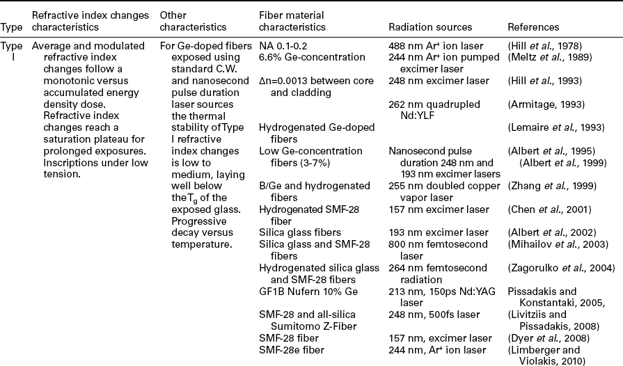

12.4.1 Type I photosensitivity
Type I photosensitive gratings are the most common category, being fabricated in low and medium concentration Ge concentration fibers under modest exposure intensities, using either CW or pulsed (Albert et al., 1995) laser sources. Their refractive index changes evolution during recording and follows a monotonic power law trend, while usually reaching saturation after the defect states have been exhausted for the single-photon absorption process; multiphoton absorption inscriptions in Ge-doped fiber can also be of Type I under controlled intensities for avoiding volume damage effects. Hydrogenated germanosilicate (Lemaire et al., 1993) fibers under low accumulated energy dose exposures also follow a Type I photosensitivity evolution. Pristine and hydrogen loaded, all-silica fibers, under 193 nm excimer laser exposure or ultraviolet and infrared femtosecond laser irradiation can also exhibit Type I photosensitivity changes (Albert et al., 2002; Smelser et al., 2005; Zagorulko et al., 2004). Type I refractive index changes are usually associated with a single underlying photosensitivity mechanism (Othonos and Kalli, 1999), or with co-occurring photosensitivity processes that contribute to the overall refractive index changes under the same sign.
12.4.2 Type II photosensitivity
Volume damage gratings induced under extreme exposure conditions by means of high energy densities (i.e. ~ 1 J/cm2 at 248 nm) (Archambault et al., 1993) or intensities (several TW/cm2 at 800 nm) (Smelser et al., 2005) are classified as Type II. In such Type II photosensitivity gratings refractive index engineering is associated with phase changes induced (Archambault, 1994) as a result of the rapid heating and resolidificaton of the exposed material, which in turn induce thermal fictive effects in the exposed glass. Type II gratings were initially demonstrated as ‘single-pulse’ gratings (Archambault et al., 1993); however, similar damage modifications can be obtained under multi pulse femtosecond laser exposures (Mihailov et al., 2003). Since such phase changes are formed above the Tg or even the melting point of the exposed glass, their thermal stability is massively increased compared to the standard Type I photosensitive gratings. Type II photosensitivity has also been observed in exposures of silica blanks (Zhang et al., 2006) using sub-MHz repetition laser sources, due to heat accumulation effects.
12.4.3 Type IIA photosensitivity
The irradiation through a phase mask of non-hydrogenated, high Ge-concentration and B/Ge-co-doped optical fibers at energy densities below the phase changes threshold revealed the complex nature Type IIA photosensitivity. First indications of the behavior of such photosensitivity were presented by Xie et al. (1993); however, a clearer demonstration of Type IIA behavior was reported by Dong et al. (1996) when a B/Ge co-doped fiber was exposed to 193 nm excimer laser radiation. In Type IIA photosensitivity both the average and modulated refractive index changes probed by the Bragg reflector inscribed follow a non-monotonic trend. Initially, the modulated refractive index changes increase similar to Type I photosensitivity, while after reaching a short plateau point, follow a declining trend leading to a turning point when the Bragg grating strength minimizes or even vanishes. Upon energy dose provided to the system the grating strength grows again, reaching a final plateau of saturation. The average refractive index changes induced into the fiber red-shift in wavelength up to the turning point of the Bragg grating strength, and then become either stable or negative/blue-shifted (Dong et al., 1996; Riant and Haller, 1997). In the broader family of Type IIA photosensitivity are included most of the complex refractive index changes induced in fiber and planar waveguide samples, where underlying photosensitivity processes are manifold and negative component refractive index changes emerge (Canning et al., 1998; Wiesmann et al., 1999). Type IIA photosensitivity inscriptions in Ge-doped fibers withstand much higher temperatures than standard Type I counterparts, exhibiting minor decay up to 800°C in specific cases (Groothoff and Canning, 2004).
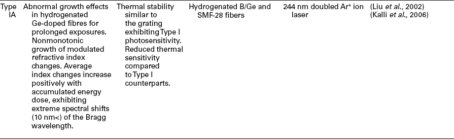
12.4.4 Type IA photosensitivity
The most recent kind of distinct photosensitivity behavior is that of Type IA, observed in hydrogenated germanosilicate fibers under prolonged exposures using CW or pulsed laser sources, however, applying intensities which can form Type IIA gratings in the same pristine fibers (Liu et al., 2002; Simpson et al., 2004). These Type IA gratings undergo significant red-shifts of the Bragg wavelength, mostly greater than 10 nm for the whole duration of the exposure. Such massive wavelength shifts correspond to average refractive index changes of the order of few parts of ×10−2, substantially modifying the guiding properties of the exposed fiber, by means of higher mode cut-off and fundamental mode confinement. The modulated refractive index behavior of Type IA gratings resembles that of Type IIA photosensitivity, where after a rapid increase the grating strength decreases to a minimum turning point, followed by a slower increase toward stabilization (Kalli et al., 2006).
12.5 Grating fabrication in standard, germanosilicate optical fibers
After the adoption of the phase mask approach (Hill, 1993; Malo et al., 1993b) as the main and most reliable Bragg grating recording method and the emergence of long-period gratings (Vengsarkar et al., 1996), the importance of understanding and simultaneously optimizing the photosensitivity processes available became quite obvious. In addition, the possibilities opened relating to commercialization of Bragg and long-period grating devices intensified the efforts in increasing germanosilicate glasses’ photosensitivity yield by means of refractive index changes and simplification of the inscription process.
There were three main axes toward the increase of the photosensitivity of germanosilicate glasses: the doping of the fiber core with ion modifiers such as boron or tin and the fiber drawing under special conditions for increasing defects and softening the glass; the hydrogen loading process; and finally the use of standard deep ultraviolet sources of photons close to the bandgap of the glass. These three approaches in combination or independently to each other, led to increase of the figure-of-merit of germanosilicate glass photosensitivity by almost three orders of magnitude from the ×10−5 levels demonstrated during the 1980s, to levels up to ×10−2. However, all these approaches included either complex processing steps, issues of repeatability and reliability or increased operational cost, rendering them less attractive for immediate commercialization. The above needs, conditions and prospects turned the research effort into alternative approaches, where the intensity and photon energy of the laser radiation used could trigger nonlinear absorption effects, bypassing the traditional single-photon photosensitivity paths. In the following subsections a review of the grating recording methods and related results will be presented focused on germanosilicate glass fibers, categorized upon the inscription wavelengths and pulse durations of the lasers used. In the last subsection the regenerated gratings will be reviewed, irrespective of recording wavelengths and pulse durations; such a type of gratings tend to constitute a new category themselves.
12.5.1 Ultraviolet nanosecond and picosecond laser inscriptions
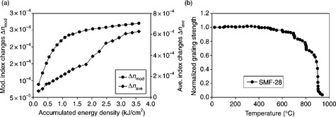
A significant demonstration of the straightforward inscription of Bragg reflectors into low-photosensitivity fibers was presented by Albert et al. (1995), after exploiting two-photon absorption effects and laser radiation of high photon energy (Fig. 12.1a). In that work, 193 nm excimer laser radiation (6.42 eV photon) was used to inscribe refractive index changes of ~ 10−3 in a hydrogen unsensitized SMF-28 standard telecom fiber, while the same irradiation produced less than half the index changes in a high Ge-doped fiber. Such refractive index changes were enough to produce Type I short and strong Bragg reflectors, in standard telecom fibers, while simultaneously avoiding the obstacles of hydrogenation. In a similar manner, Herman et al. exploited the much shorter photon wavelength of 157 nm (7.9 eV) for inscribing long-period gratings in pristine SMF-28 fibers (Chen et al., 2001), providing energy to the system above the germanosilicate core glass bandgap, while allowing single-photon ionization processing (Dyer et al., 2001) and refractive index changes of the order of ~ 4 × 10⁻⁴. Compaction was the primary underlying physical mechanism in the 157 nm exposures, while cladding absorption defined a rather narrow envelope for optimum inscription conditions. The above two deep ultraviolet wavelengths were also used for locking photosensitivity in hydrogen loaded fibers (Chen et al., 2002b), as well as for Bragg grating amplification (Dyer et al., 1994). Later, Dyer et al. (2008) used 157 nm nanosecond laser radiation and a custom-made CaF2 phase mask for recording Bragg reflectors in low-defect SMF-28 and Hi-980 Corning fibers (Fig. 12.1b), leading to refractive index changes of ~ 2.8 × 10⁻⁴, extending the work of Herman and Chen (Chen et al., 2001).

12.1 (a) Growth of refractive index modulation amplitude in Low-Ge fiber (SMF-28 Corning) resulting from irradiation through a phase mask with ArF laser at 50 pulses/s with pulses of different energy density (after Albert et al., 1995). (b) Comparison of spectral reflectivity of FBG in HI-980 and SMF-28 fiber versus 157 nm laser dose. Fluence per pulse ~ 58 mJ/cm2. The solid line represents the modulation amplitude of refractive index for SMF 28 fiber. (After Dyer et al., 2008. Used with permission from Optical Society of America [OSA].)
In 2005 there was presented the first inscription of Bragg reflectors in a germanosilicate fiber using 213 nm, 150 ps frequency quintupled Nd:YAG laser radiation, targeting the low absorption spectral valley formed between the Ge oxygen deficiency centers peaking at 5 eV, and the GeE’ centers absorption band tail initiated at this wavelength regime (Pissadakis and Konstantaki, 2005b). Spectro-photometric measurements of 9% mole Ge-doped fiber performs presented by Archambault (1994) reveal that the absorption at the valley of the 213 nm wavelength is more than 10× lower that that measured at the 242 nm germanium-oxygen deficiency band, heavily exploited in grating recording using 248 nm excimer lasers.
Average index changes Δn ≈ 8.5 × 10⁻⁴ were obtained after 2 hours’ exposure of the Nufern GF1B fiber with a cumulative energy density of 2.9 kJ/cm2 (Fig. 12.2a and 12.2b), while employing an elliptical Talbot interferometer (Pissadakis and Reekie, 2005). The Talbot interferometer configuration imposed adequate separation between the same mask and the fiber, allowing high energy density delivery in the fiber complex, without significant absorption in the phase mask due to the SiE’ centers formation located at the 5.8 eV band (Skuja, 1998). The refractive index growth data followed the universal power law trend applied in the case of compaction in silicate glasses, exhibiting a b factor of ~ 0.66 (Borrelli et al., 1999; Schenker, 1994), described by Eq. [12.3]. The GW/cm2 magnitude intensities used in the inscription (Pissadakis and Konstantaki, 2005b) in conjunction with the high two-photon absorption coefficient of the germanosilicate core led to a nonlinear absorption coefficient similar or even higher than the linear absorption at this wavelength (Kalachev et al., 2005), promoting two-photon effects. Exposures performed using different energy densities in the Nufern GF1B fiber support further the above assertion, by leading to refractive index changes such that their ratio is proportional to the second power of the corresponding ratio of the energy densities per pulse (see Fig. 12.3).
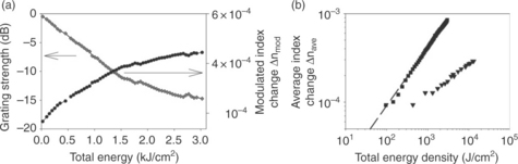
12.2 (a) Bragg grating strength (black cycles) and modulated refractive index changes Δnmod (gray diamonds) versus total energy density for Nufern GF1B fiber using 213 nm Nd:YAG laser radiation, using 42 mJ/cm2 energy density per pulse. (b) Comparative graph on the average refractive index changes Δnave versus total energy density for Bragg grating recording using 213 nm, 150 ps 213 nm Nd:YAG (black squares) and 248 nm, 34 ns excimer (inverse triangles) laser radiation. Energy density for 248 nm exposure: 360 mJ/cm2. Dashed line: power law regression using Eq. [12.3] for average refractive index changes induced by 213 nm laser radiation.

12.3 Average refractive index data Δnave for Bragg gratings inscribed in Nufern GF1B optical fiber using 213 nm, 150 ps Nd:YAG laser radiation, for different energy densities per pulse. Triangles: 42 mJ/cm2. Circles: 35 mJ/cm2. The vertical line defines the iso-energy point of ~ 1.2 kJ/cm2, where the two gratings are measured.
Using 213 nm wavelength and 150 ps pulse duration the recording of Type IIA Bragg reflectors in the commercial PS1250/1500 Fibercore B/Ge co-doped fiber was also presented, leading to refractive index changes of 9.0 × 10⁻⁴ (Pissadakis and Konstantaki, 2005a). Again, the refractive index changes data referring to the Type IIA evolution regime were well fitted to a power law growth (see Eq. [12.3]), with b factor 0.65 (Fig. 12.4).

12.4 Index modulation Δnmod versus accumulated energy, for a Type IIA Bragg grating exposure in the B/Ge-codoped PS1250/1500 Fibercore optical fiber, of 72 000 pulses and a pulse energy density of 60 mJ/cm2.
Both the results obtained for the Nufern GF1B and the PS1250/1500 Fibercore optical fibers during Bragg grating inscriptions using 213 nm, 150 ps Nd:YAG radiation, provide evidence that the primary underlying physical mechanism was that of volume compaction. The first surprising finding that supports the compaction model emerges from the power law fitting of the refractive index data for both fibers used and exposed using the 213 nm, 150 ps. Both fibers exhibit almost identical b-factor value after fitting their photosensitivity data using Eq. [12.3]; similar b-values have been also reported by Borrelli (Borrelli et al., 1999) and Schenker (1994). Another experimental finding in support of the compaction model for the 213 nm, 150 ps irradiation is obtained by producing Bragg reflectors using 248 nm, nanosecond excimer laser radiation, utilizing extreme energy densities (~ 1 J/cm2 per pulse). By comparing the data for the 213 and 248 nm exposures under the dose conditions of Fig. 12.5, one can see that initially the 248 nm exposure exhausts 242 nm peaking germanium-oxygen deficiency centers and their corresponding refractive index contribution; then follows an identical growth trend with the 213 nm exposure. Since the 213 nm, 150 ps radiation does not primarily rely on the exhausting of color centers for inducing refractive index changes, both laser wavelengths produce the same type of changes in the glass (Pissadakis and Konstantaki, 2005b), after specific dose threshold has been surpassed.

12.5 Average refractive index data Δnave versus radiation dose for comparative Bragg grating exposures using 213 nm, 150 ps (circles) and 248 nm, 34 ns (triangles) laser radiation.
Shortly after the above reports on 213 nm photosensitivity, 211 nm 250 fs radiation was used for long-period grating recording in hydrogenated SMF-28 and B/Ge co-doped fibers, following a clear Type I photosensitivity behavior for both fibers exposed (Kalachev et al., 2005). Recently 30 KHz, 213 nm Nd:VO4, 7 ns laser radiation was used for recording Bragg ratings in hydrogen-free, SMF-28 fiber, achieving refractive index changes of the order of 10−3 (Gagné and Kashyap, 2010). Gagné and Kashyap supported that the primary physical mechanism dominating the photosensitivity of SMF-28 fiber using 213 nm nanosecond radiation was of a single-photon absorption nature, prompting color-center generation. The single-photon color-center generation was associated with the longer pulse duration employed.
12.5.2 Infrared femtosecond laser inscriptions
There have been early investigations on the use of femtosecond laser radiation in the refractive index engineering of optical fibers, starting from the work of Saifi et al. (1989), who measured permanent refractive index changes of few parts of ×10−5 induced in a twin core, germanosilicate optical fiber, directional coupler by exposure to 620 nm, 100 fs laser radiation. Saifi et al. quantified these changes by measuring changes in the beat-length of the directional coupler, by power coupling between the two cores, while attributing for the first time the photosensitivity obtained in multiphoton generated structural changes (Griscom, 2011). A decade later, Cho et al. (1999) observed increase of the SiE’ centers after the formation of plasma channelling in a multimode silica fiber using 790 nm, 110 fs Ti:Sapphire laser radiation. The formation of plasma channelling induced a permanent double cladding structure into the multimode silica fiber of 2 × 10⁻² refractive index contrast. Then, Fertein et al. (2001) measured 6 × 10⁻³ refractive index changes using in the cavity length a Bragg gratings Fabry-Perot in Corning fiber SMF-28, exposed using 800 nm femtosecond laser, at a focal spot of 0.4 mm.
The landscape of fiber photosensitivity and grating recording was redrawn after the first Bragg reflectors inscribed in SMF-28 and all-silica depressed cladding optical fibers by Mihailov et al. (2003, 2004). In this work, both SMF-28 and all-silica fibers were exposed using a modified design phase mask for forming short length and highly scattering Bragg reflectors (Fig. 12.6), which maintained their reflectivities in temperatures greater than 1000°C.

12.6 (a) Reflectivity of a high-order grating written in a SMF-28 fiber with the 2.142 μm pitch mask and 800 nm and 120 fs Ti:Sapphire laser radiation. (After Mihailov et al., 2003. Used with permission from Optical Society of America [OSA].) (b) Variation of the reflectivity and resonant wavelength of a grating recorded using 800 nm and 120 fs Ti:Sapphire laser radiation with the number of incident IR pulses (300 mJ pulse, 10 Hz). The squares denote reflectivity; circles, wavelength shift. (After Mihailov et al., 2004. Used with permission from IEEE.)
The refractive index changes were a product of a four/five-photons (depending upon the material bandgap) nonlinear absorption at intensities of 1.2 × 10¹³ W/cm2. That group exposed hydrogenated SMF-28 fibers using the same setup and conditions revealing reduction of compaction effects in the silica glass cladding (Limberger et al., 2007), while the gratings exhibited annealing behavior similar to those inscribed using ultraviolet radiation (Smelser et al., 2004). By varying exposure energy densities pure Type I-IR or Type II-IR gratings could be formed in SMF-28 fibers, where Type I-IR are possibly associated with color-center accumulation, and Type II-IR are products of extreme ionization and plasma formation processes. Type I, ultrabroad bandwidth, chirped Bragg reflectors were fabricated using Ti:Sapphire 800 nm, 1 KHz femtosecond laser radiation, in hydrogenated and pristine standard telecom fibers, using highly chirped phase masks, achieving FWHM bandwidths greater than 200 nm (Bernier et al., 2009).
Alternatively to the use of custom design phase masks for controlling spatial dispersion and beam de-condensation effects that can detrimentally affect the high intensities required during femtosecond laser refractive index engineering (Mihailov et al., 2004), point-by-point Bragg grating recordings were performed in standard telecom fibers (Martinez et al., 2004). The nonlinear refractive index engineering at sub-wavelength volumes using 800 nm femtosecond lasers (Glezer et al., 1996) offers significant advantages in the point-by-point Bragg grating processing, allowing ease in tailoring the topology of the periodic structure as well as its polarization characteristics. Martinez et al. inscribed such reflectors operating in the first diffraction and also in higher orders, using high magnification objectives and an 800 nm, 1 kHz, 150 fs Ti:Sapphire laser (Fig. 12.7). The Bragg reflectors inscribed were almost 25 dB in strength, exhibiting typical birefringence of ~ 3 × 10⁻⁵ due to the highly asymmetrical nature of the inscription process. Using the same method Martinez et al. (2006) also presented Bragg grating inscription in a jacket unstripped standard telecom fiber, succeeding in grating strengths greater than 25 dB. The above was a significant improvement compared to the previous art, where special coatings were used for inscribing Bragg reflectors through the fiber jackets using 244 nm laser radiation (Chao et al., 1999). Infrared femtosecond laser point-by-point Bragg grating inscription technique was also used for recording apodized geometry structures in SMF-28e fibers by adopting a slanted scanning technique with respect to the fiber core (Williams et al., 2011b) or complex sampled and phase-shifted Bragg reflectors (Marshall et al., 2010).

12.7 Transmission spectra measured in gratings of first, second and third orders fabricated using 800 nm femtosecond radiation and point-by-point technique. (After Martinez et al., 2004. IET copyright permission is acknowledged.)
Different kinds of Bragg reflectors were inscribed in standard telecom fibers exploiting filamentation effects, induced under Ti:Sapphire 800 nm femtosecond laser radiation (Bernier et al., 2011). High energy density inscriptions can lead to filamentation generation and propagation inside the fiber complex, inducing refractive index changes as high as ×10−2 (Sudrie et al., 2002; Yamada et al., 2001). Due to the nature of filament generation and propagation, the modifications induced in the hosting glass matrix are spatially localized in typical dimensions below a few microns, allowing high refractive index contrasts, thus increased diffraction efficiencies. Bernier et al. (2011) reported grating inscription by cross-sectioning the fiber core with the periodic filament generated by a phase mask, inducing refractive index changes of ~ 2.5 × 10⁻³.
12.5.3 Ultraviolet femtosecond laser inscriptions
While in IR femtosecond exposures several photons were required for covering the large bandgap of germanosilicate glasses and fused silica, at the expense of tight focusing and narrow envelope inscription conditions, the use of ultraviolet picoseconds and femtosecond sources could alleviate these tight inscription conditions exploiting lower-order, nonlinear effects. The scaling down rule of the damage fluence threshold of an optical material versus pulse duration (Boyd, 2003; Stuart et al., 1995) applies also in photosensitivity, questioning the efficiency of traditional laser wavelengths such as 248 and 266 nm into the inscription of higher index changes at lower energy doses. The first inscriptions of Bragg gratings in hydrogenated SMF-28 fibers were presented using a low repetition rate, frequency quadrupled 264 nm Nd:glass laser (Dragomir et al., 2003), at maximum intensities of 77 GW/cm2.
Comparative results on the Bragg grating inscription process in SMF-28, phosphorus doped and all-silica fibers using 1 KHz repetition rate, tripled 267 nm, Ti:Sapphire femtosecond laser with results obtained using 157 and 193 nm ultraviolet excimer lasers (Zagorulko et al., 2004). Zagorulko et al.’s study revealed that the photosensitivity of hydrogenated, low-Ge content silicate glass fibers using femtosecond, ultraviolet laser resembles that obtained using 157 nm excimer laser, leading to similar refractive index changes (few parts of 10−3) and growth rates, while suffering less from saturation effects (Fig. 12.8).

12.8 Dependence of refractive-index changes on exposure dose of an H2-loaded SMF-28 fiber for inscriptions using 267, 157 and 193 nm laser wavelengths and corresponding pulse durations. (After Zagorulko et al., 2004. Used with permission from Optical Society of America [OSA].)
The fabrication of long-period gratings in hydrogenated SMF-28 fiber variants using 352 nm, emitted from a frequency tripled Nd:glass laser and point-by-point exposure, was reported by Dubov et al. (2005). In this report a three-photon absorption process was triggered at intensities of the order up to 2000 GW/cm2, while cladding damage was visible denoting asymmetrical absorption across the fiber cross-section.
Strong Bragg reflectors were also fabricated in SMF-28 fibers for modest accumulated energy densities (Livitziis and Pissadakis, 2008), using a double phase mask interferometer and 248 nm, 500 fs laser radiation (see Fig. 12.9a). These Type I gratings saturated at accumulated energy densities as low as 3.5 kJ/cm2, reaching refractive index changes up to ~ 7 × 10⁻⁴, while exhibiting thermal durability up to 900°C (Fig. 12.9b). The enhanced thermal stability of these SMF-28 fiber gratings, compared to those recorded using 193 nm excimer laser radiation (Albert et al., 1995), was associated with the higher two-photon absorption coefficient of the Ge-doped core (Dragomir, 2002), which in turn may lead to a substantial increase of the local temperature levels in the bright fringes.
12.9 (a) Refractive index modulation Δnmod (circles) and average index Δnave (diamonds) changes versus accumulated energy density for grating exposure in SMF-28 optical fiber using 248 nm, 500 fs laser radiation. (b) Isochronal thermal annealing results for a Bragg grating recorded in SMF-28 optical fiber, using 248 nm, 500 fs laser radiation.
Bragg and long-period grating inscriptions using femtosecond lasers produced mostly Type I and Type II photosensitive refractive index changes, while no Type IIA had been produced either by 800 nm Ti:Sapphire or quadrupled Nd:glass lasers, for prolonged irradiations and energy density doses into germanosilicate glasses fibers irrespective of Ge concentration and codopands. The first Type IIA Bragg grating recording utilizing femtosecond laser radiation was presented by Violakis et al. (2006) using 248 nm, 500 fs hybrid excimer/dye laser and phase mask configuration in contact mode. The fibers exposed were the B/Ge co-doped, PS1250/1500 from Fibrecore and a Hi-Ge from FiberLogix (Fig. 12.10a,b).
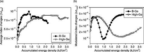
12.10 (a) Index modulation Δnmod and (b) average index Δnave changes versus accumulated energy density, for Type IIA Bragg grating exposure of B-Ge and High-Ge optical fibers using 248 nm, 500 fs excimer laser radiation.
Violakis et al. presented comparative data with similar inscriptions using 248 nm, 34 ns excimer laser radiation, where the femtosecond grating recording was saturated for doses 10× times smaller than those required in the nanosecond recording; while both Type I and Type IIA refractive index changes slopes were accelerated for the femtosecond exposure case (Fig. 12.11). In addition, annealing studies revealed that the Type IIA gratings recorded using 248 nm, 500 fs radiation demarcated at 700°C, 100°C higher than the nanosecond counterparts, due to structural changes formed by the irradiation (Violakis et al., 2006).

12.11 Comparative graph for index modulation Δnmod changes recorded for grating inscription in PS1250/1500 B-Ge fiber using 248 nm, 500 fs and 34 ns excimer laser radiation.
In later studies performed by the same group (Pissadakis et al., 2006), comparative Type IIA Bragg grating recordings were demonstrated using 5 ps, 500 and 120 fs, 248 nm radiation together with micro-Raman studies, investigating the effect of exposure conditions in the photosensitivity growth characteristics, by performing exposures for fixed intensity and energy density and different pulse durations (Fig. 12.12a). Exposures performed under fixed intensities for different pulse durations illustrated that the effect of the energy density plays a predominant role in the triggering of the Type IIA photosensitivity mechanism, while intensity rather affects its progression speed (Fig. 12.12b).

12.12 (a) Index modulation Δnmod changes versus accumulated fluence, for Bragg grating exposure in B-Ge optical fiber using 248 nm, 120 fs, 500 fs and 5 ps laser radiation, of fixed fluence. (b) Index modulation Δnmod changes versus accumulated fluence, for grating exposures of fixed intensity in the same optical fiber using 120 and 500 fs laser pulse duration.
Additionally, the 248 nm femtosecond laser irradiation modifies the characteristics of the Boson peak (Hehlen et al., 2002) in Raman spectra obtained (Dianov et al., 1997b) from the boron-germanosilicate glass core, shifting this to longer wave numbers (Fig. 12.13), while exhibiting similarities to the response of silicate glasses subjected to hydrostatic pressure above the plasticity level (Inamura et al., 1997).
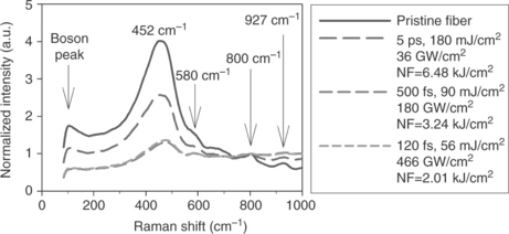
12.13 Micro-Raman scattering spectra for unexposed and exposed B-Ge doped fiber cores, using different pulse durations, for isochronal exposures using N = 36 000 pulses. NF: total accumulated energy fluence in J/cm2. The micro-Raman scattering spectra were obtained using 473 nm line of an Ar+-ion laser, while they have been normalized to the strength of the Si-O 800 cm−1 peak.
The modification of the Boson peak constitutes a strong indication that the primary underlying mechanism behind Type IIA photosensitivity is that of compaction in the bright fringes of interference, and secondary that of stress relief; that was also described by Ky et al. (2003b).
12.5.4 Regenerated gratings
Bragg grating regeneration and post-exposure amplification were first observed in Type IIA gratings fabricated using 193 nm, excimer laser radiation in a B/Ge co-doped optical fiber (Dong and Liu, 1997). In such grating type, the exposure was ceased before reaching Type IIA saturation, and then the fiber reflector was subjected to annealing processes, where amplification strength was observed. Similar abnormal thermal annealing behavior had also been observed in Bragg gratings fabricated in OH-flooded, F-depressed cladding fiber at temperatures within the range of 1000°C (Fokine, 2002a), attributed to molecular water formation and diffusion into the silica glass matrix, and subsequent oxygen reduction. Then, thermal regeneration was also observed in a hydrogenated, F, P and B/Ge co-doped fiber with high concentration of both B and Ge ions that had been exposed to 193 nm, for forming a Type I Bragg reflector (Bandyopadhyay et al., 2008).
Canning et al. (Bandyopadhyay et al., 2008) speculate that the grating writing process leaves a structural signature in the exposed glass matrix, which is not demarked by the thermal treatment, even though the refractive index changes may erase. Instead such structural signature imprinted in the glass may constitute a catalytic base for assisting chemical reactions with hydrogen for forming possible species of hydrite or hydroxyl groups that can induce local stresses in the exposed fringe level (Canning et al., 2008b). Further, the above regenerated gratings exhibited extreme durability to thermal treatment, withstanding temperatures up to 1000°C (Fig. 12.14). Similar regeneration results were obtained for gratings fabricated in high Ge content fibers under strained annealing (Lindner et al., 2011), while surviving under similar temperature conditions as those presented in (Bandyopadhyay et al., 2008).
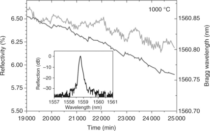
12.14 Annealing behavior of refractive index modulation and Bragg wavelength of regenerated grating after 2 weeks at 1000 °C. Inset: grating spectrum (Lindner et al., 2011).
Thermal regeneration behavior has been observed for Type IIA Bragg gratings fabricated using femtosecond and picosecond 248 nm laser radiation (Pissadakis et al., 2006), for exposures ceased before reaching the Type IIA saturation level (Fig. 12.15).

12.15 (a) Index modulation Δnmod changes versus accumulated energy density for Bragg grating exposure in B-Ge optical fiber using 248 nm, 120 fs, 500 fs and 5 ps laser radiation, of similar fluence. (b) Normalized grating strength, during regeneration annealing at isothermal temperature steps of 100 °C, of the previous Bragg gratings until erasure point is reached.
Figure 12.15a shows index modulation Δnmod changes versus accumulated energy density for Bragg grating exposure in B-Ge optical fiber using 248 nm, 120 fs, 500 fs and 5 ps laser radiation, of similar fluence. Figure 12.15b shows normalized grating strength, during regeneration annealing at isothermal temperature steps of 100°C, of the previous Bragg gratings until erasure point is reached.
Three different Bragg gratings exposures performed in the PS1250/1500 Fibercore B/Ge codoped fiber using 5 ps, 500 fs and 120 fs while keeping similar energy densities, ceased when reaching the same refractive index change level at the Type IIA photosensitivity regime. Accordingly all gratings were annealed from room temperature up to 600°C, reaching full demarcation. These three gratings exhibited almost identical regeneration characteristics, amplifying in strength with increasing temperature, reaching the maximum strength point at 550°C approximately; then erasing until the temperature of 600°C. Such behavior reveals that the glass composition and thermal history dominates the specific regeneration process, while the exposure conditions play rather a secondary role. Therefore, the regeneration characteristics may be tuned by changing the pre-exposure thermal history of the glass (i.e. rapid heating and cooling process), for modifying the population and type of fictive defects in the matrix, that can be later seeded by the irradiation process (Bandyopadhyay et al., 2008).
12.6 Grating fabrication in standard, all-silica optical fibers
While in germanosilicate glass fibers there was significant progress related to grating inscription and refractive index engineering, the formation of photosensitive refractive index changes in all-silica fibers remained a rather hard task, thus frustrating the optimum exploitation of those fibers in photonic devices development due to lack of inscription processes. The photosensitivity of the high quality silicate glass at the spectral band from 5 to 8 eV is dependent upon low concentration of defects such as oxygen deficiencies, Si-Si wrong bonds and SiE’ centers peaking at 5.8 eV, with significant tail up to 6.5 eV (Skuja, 1998). The state-of-the-art was limited to irradiations using 193 nm excimer laser and several hundred thousand pulses for inducing refractive index changes of the order of 10−5 (Rothschild et al., 1989); being insufficient for forming strong Bragg reflectors in allsilica fibers.
The formation of the first strong Bragg reflectors in depressed cladding all-silica fibers using 193 nm laser radiation (Albert et al., 2002) that have been subjected to hydrogen flooding and OH defect generation (Lancry et al., 2007), was the first demonstration of photo-engineering of refractive index (Δn ~ 0.5 × 10⁻⁴) in such fibers (Fig. 12.16). The formation mechanism of those reflectors was classified as of chemical nature where Si-OH species initially formed by the high temperature flooding are photo-activated for forming water species in the bright fringes of interference, while simultaneously inducing photorefractive effects and stress generation into the matrix (Fokine, 2002a; Smith et al., 2001). In the work of Albert et al. pristine F-depressed cladding fiber was exposed, producing Bragg grating reflectors of smaller diffraction efficiency than the OH-flooded one, with corresponding refractive index changes of few parts of ×10−5. Shortly after the demonstration of Albert and Fokine (Albert et al., 2002), strong Bragg reflectors were recorded in non-sensitized silica glass, optical fibers by Mihailov et al. (2004) employing a 800 nm femtosecond Ti:Sapphire laser, expanding the tools available for refractive index engineering of all-silica glass fibers.

12.16 Growth in refractive-index modulation for deuterium-loaded and pristine all-silica, fluorine depressed cladding fibers. (After Albert et al., 2002. Used with permission from Optical Society of America [OSA].)
Since the Bragg grating fabrication in all-silica fibers was successful using 800 nm, a fundamental question arose relating to the combination of ultraviolet radiation with photon energy comparable to the bandgap of the silica glass with femtosecond pulse duration for improving further the recording yield. In such a combination the order of multiphoton absorption would lower to 2–3 photons, alleviating focusing requirements, while accessing centers closer to the Urbach tail of the glass (Kühnlenz et al., 2000). First Bragg gratings inscription attempts in pristine, all-silica fibers were presented in (Zagorulko et al., 2004) using frequency tripled 800 nm Ti:Sapphire laser emitting at 267 nm, while using a phase mask in contact mode. However, in this work the yield of Bragg grating inscription in the non-hydrogenated silica glass fiber was low, forming reflectors less than 0.1 dB in strength after accumulated energy density doses of ~ 50 kJ/cm2. This may be associated with the photon energy of 267 nm which is ~ 4.6 eV marginally above the nominal bandgap of silica glass if considered in a two-photon absorption scheme (Dragonmir et al., 2002).
By adopting slightly shorter wavelength at 248 nm and a substantially different inscription setup, strong Bragg gratings were inscribed in hydrogen unloaded, low-OH concentration, all-silica Sumitomo Z-fiber by Livitziis et al. (Livitziis and Pissadakis, 2008), using 500 fs laser pulses. Livitziis et al. used a dual phase mask interferometer detaching the phase mask element from the fiber (Fig. 12.17a) and allowing the delivery of sub-TW/cm2 intensities in the fiber complex, without suffering from two-photon absorption and color-center accumulation in the phase mask (Taylor et al., 1988).
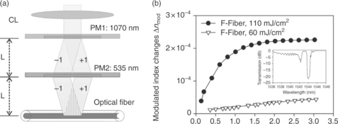
12.17 (a) Schematic diagram of the double phase mask interferometer. CL: cylindrical lens. PM1, 2: phase mask elements with periodicity annotated. (b) Refractive index modulation Δnmod changes versus accumulated energy density for grating exposures in all-silica Sumitomo Z-fiber, using 248 nm, 500 fs laser radiation, for two different energy densities. Inset: transmission spectrum of the Bragg reflector inscribed using 110 mJ/cm2 energy density.
The combination of the 248 nm wavelength (of ~ 5 eV photon energy) and the extreme intensities delivered in the fiber, boosted two-photon absorption effects, and led to the formation of modulated refractive index changes of the order of ~ 2.2 × 10⁻⁴ (and of 5 × 10⁻⁴ average refractive index changes) for intensities of (≈ 440 GW/cm2) at the bright fringes of the interference pattern (Fig. 12.17b). Inscriptions using different energy densities verify the two-photon absorption nature of the inscription process, where the refractive index changes obtained are dependent upon the second power of the inscription intensity. Also, the appearance of low cladding mode notches in the transmission spectra of the gratings recorded using 248 nm, 500 fs laser radiation, indicates that refractive index changes have also been inscribed in the radiation resistant fluorine doped cladding (Hosono, 1999).
A useful comparison of the trend and the yield of the 248 nm, 500 fs inscription, with one performed using 193 nm, 10 ns laser radiation in the same all-silica fiber, presented in Fig. 12.18, illustrates that the photosensitivity processes induced by these two wavelengths are different (Fig. 12.18a). The two-photon mediated photosensitivity process of the 248 nm, 500 fs radiation progresses faster with respect to energy density dissipated in the fiber; the intermediate germanium-oxygen deficiencies state at 5 eV can also assist such accelerated behavior. The corresponding exposure using 193 nm radiation exhibits different slope shape; the defects accessed are possibly of a broader nature close to the band-gap of the material. However, subsequent annealing of the Bragg reflectors produced using those lasers and pulse durations denotes that the type of modifications produced are of the same type (Fig. 12.18b).
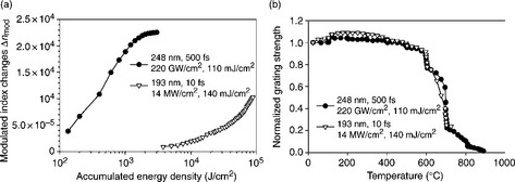
12.18 (a) Refractive index modulation Δnmod changes versus accumulated energy density for grating exposures in all-silica Sumitomo Z-fiber, using 248 nm, 500 fs and 193 nm, 10 ns laser radiation. (b) Annealing study of the reflectors produced using the above wavelengths and pulse durations.
A standard Talbot interferometer was used for recording Bragg reflectors inside a Al/Yb co-doped all-silica standard fiber using 262 nm femtosecond laser pulses, emitted by a frequency tripled Ti:Sapphire laser (Becker et al., 2008). This report re confirmed that by increasing the spacing between the phase mask and the fiber energy dissipation constraints are alleviated, leading to efficient inscription inside all-silica glass fibers using ultraviolet femtosecond lasers. Long exposures of energy doses greater than 2 MJ/cm2, using CW frequency doubled Ar+ ion laser radiation were used for forming Bragg reflectors in unsensitized Ge-free, Bi-Al, silica glass fibers (Violakis et al., 2011), reaching refractive index changes up to 3.5 × 10⁻⁴. The possible mechanism of grating formation using such continuous wave (CW) exposures can be two-photon absorption-related compaction, progressing at slow rates due to the lack of sufficient intensities; pre-existing color centers exhausting are saturated during the early phases of inscription.
Other interesting results included the Bragg grating inscription in N-doped core silica-glass fibers using 193 nm laser radiation and standard phase mask setup (Butov et al., 2006; Dianov et al., 1997a), where a clear Type IIA photosensitivity behavior was monitored during inscription (Fig. 12.19a). The Type IIA gratings recorded in the N-doped fiber exhibited thermal regeneration characteristics, withstanding temperatures up to 1200°C (Fig. 12.19b). The authors of this work (Butov et al., 2006) justify the extreme thermal stability and regeneration effects upon a nitrogen-species mobility model, where the photo-excited nitrogen can diffuse and be re-trapped from the core to the cladding area forming structural corrugations. In later work on N-doped silica fibers gratings recorded using 193 nm excimer laser, Lanin et al. (2007) observed similar thermal and non-thermal regeneration effects, of non-reversible nature, upon hydrogen diffusion into the pre-exposed fiber, due to thermochemical reaction of hydrogen toward the formation of Si-NH and Si-OH species.

12.19 (a) Effective refractive index modulation in a Bragg grating (λB = 1540 nm) as a function of exposure dose of a 193 nm wavelength laser radiation, the power density per pulse being 100 mJ/cm2, pulse duration 8ns and repetition rate 100 Hz. (b) Isochronal annealing of Type IIA Bragg gratings written in an N-doped fiber under different regimes. (After Butov et al., 2006.)
12.7 Grating fabrication in phosphate and fluoride glass fibers
12.7.1 Phosphate glass fiber gratings
The photosensitivity of phosphorus codoped silicate glass fibers has been investigated since the 1990s, when 193 and 240 nm pulsed laser radiation was used for inducing transient and permanent refractive index in those fibers (Canning et al., 1995). The permanent residue of refractive index changes obtained for hydrogen loaded, phosphorus doped silica glasses was of the order of 10−3, under 193 nm irradiation; unloaded fibers were quite less photosensitive. It was also known that phosphorus dopand suppresses the 242 nm germanium-oxygen deficiencies band when inserted in germanosilicate glasses, acting as a passivator for such types of wrong bonds, thus reducing photosensitivity (Dong et al., 1994).
The valence state of phosphorus allowing the formation of single and double bonds with oxygen, defines the micro-coordination of phosphate glasses while leading to a linear-like polymerization chain built in combination with oxygen and other ion modifiers; a micro-coordination structure which is substantially different from that of silicate glasses. Due to this versatile phosphorus–oxygen bond state, phosphate glasses exhibit interesting optical, chemical and mechanical properties, dependent upon the ratio between phosphorus and oxygen ion concentrations, and the incorporation of other matrix modifiers. Phosphate glasses are highly transparent and durable to ultraviolet radiation, have a high solubility of rare-earth ions and exhibit low Tg points (Ehrt et al., 1994). Rare-earth doped phosphate glasses have been drawn into standard and MOFs and planar waveguides for realizing high gain amplifiers (Hwang et al., 1999) and lasers (Li et al., 2005) of short physical lengths. Therefore, the straightforward inscription of Bragg reflectors in those guiding structures is required for realizing lasing devices and sensors (Strasser, 1996). The first studies of the photosensitivity of pure phosphate glasses were presented in the mid-1990s, mostly focused on fluorophosphates slab samples including Ce+ ion modifiers while being irradiated using 248 and 193 nm excimer laser radiation (Ebendorff-Heidepriem and Ehrt, 1996; Ebendorff-Heidepriem et al., 2002). In these studies spectroscopic modifications were solely examined, rather than refractive index engineering.
Generally, the exposure of phosphate glasses to ultraviolet laser and X-ray radiation results in color-center generation extended from the visible to the ultraviolet and bandgap spectral regime, associated with the transformation of the phosphorus–oxygen bond and the related defects, with most prominent the PO hole center in the visible band and the PO4 electron center peaking at the 242 nm wavelength (Ebeling et al., 2002; Ehrt et al., 2000). Further to these first photosensitivity studies carried out using short wavelength sources and single-photon absorption excitation, there were presented refractive index engineering demonstrations using infrared (Chan et al., 2003) and near ultraviolet femtosecond lasers in pristine and silver doped phosphate glass substrates (Watanabe, 2001), respectively.
A study focusing on the refractive index engineering of the commercially available rare-earth doped IOG-1 Schott glass including the irradiation of pristine and silver ion-exchanged samples using 248 nm nanosecond excimer laser was presented in 2004 (Pissadakis et al., 2004). This study revealed that the refractive index changes obtained in a pristine glass slab for accumulated energy density doses of 12 kJ/cm2 was of the order of a few parts of ×10−5, while the addition of silver boosted the photosensitivity of the glass by almost three orders of magnitude. The same glass exposed using 213 nm, 150 ps Nd:YAG laser and an elliptical Talbot interferometer exhibited similar refractive index changes (few parts ×10−5); however, non-monotonic growth effects were monitored during grating inscription (Pappas and Pissadakis, 2006).
Other investigations in the same IOG-1 glass using 193 nm laser radiation led to greater photosensitivity effects that were directly exploited for waveguide laser development (Yliniemi et al., 2006a, 2006b). Most importantly the work of Yliniemi et al. revealed that phosphate glass undergoes structural changes under 193 nm laser irradiation including volume modification, confirmed by utilizing micro-Raman spectroscopy and atomic force microscopy. Similar findings related to volume modifications induced by 193 nm (Michelakaki and Pissadakis, 2009) and 248 nm, 500 fs (Pissadakis and Michelakaki, 2008) laser radiation were presented by Michelekaki et al. where Knoop micro-hardness measurements were used for monitoring non-monotonic volume dilation effects versus energy density dose, under a single-photon absorption mechanism (Fig. 12.20a).
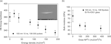
12.20 Knoop hardness versus energy density of IOG1 phosphate glass exposed using 193 nm, 10 ns excimer laser radiation. Inset: Knoop hardness indentation imprint on a phosphate glass sample. Elastic modulus versus radiation dose of IOG1 glass exposed using 193 nm, 10 ns excimer laser radiation. The phosphate glass for the exposure conditions above undergoes volume dilation.
From the variation of the Knoop hardness data obtained for 193 nm excimer laser exposures in bulk phosphate glass samples, the corresponding changes in the elastic modulus were evaluated, confirming the volume dilation induced by the irradiation process (Fig. 12.20b). Such atypical radiation-induced volume modification effects were attributed to PO bond transformation from single to double and subsequent cleaving upon the conditions of the irradiation process.
The aforementioned investigations on the photosensitivity of bulk phosphate glass samples constituted a solid background for attempting Bragg grating inscription in all-phosphate glass fibers. The first Bragg gratings in a rare-earth doped all-phosphate glass fiber were demonstrated by Albert et al. (2006) using 193 nm excimer laser and standard phase mask inscription technique. Albert et al. inscribed strong, Type I Bragg reflectors in a phosphate glass fiber, obtaining average refractive index changes of 5 × 10⁻⁴. These gratings under low temperature annealing, amplified more than 65% in strength; this behavior was attributed to slow stress relaxation effects accelerated by the thermal treatment (Albert et al., 2006; Rodica Matei et al., 2007). Grobnic et al. (2007) followed with positive refractive index, Type I Bragg grating inscriptions in a similar phosphate glass fiber using 800 nm, femtosecond laser radiation, reaching refractive index changes greater than 1.5 × 10⁻³. These reflectors exhibited rather standard thermal stability, without post-exposure amplification effects, while maintaining their strengths up to temperatures of 400°C. Notably, the results of Grobnic et al. (2007) oppose earlier observations of Chan et al. (2003) where negative refractive index changes formed waveguides in IOG-1 glass under 800 nm, femtosecond irradiation; these negative refractive index changes demarcated at substantially lower temperatures.
Recently, Sozzi et al. exposed the same fiber as Grobnic et al. (2007) using 248 nm, 500 fs laser radiation and a double phase mask interferometer (Sozzi et al., 2011). Sozzi et al. found that at high energy densities for such pulse duration, a photosensitivity mechanism similar to Type IIA was activated in the phosphate glass fiber, following non-monotonic growth of both the average and modulated refractive index changes (Fig. 12.21a). The last was the first demonstration of Type IIA-like photosensitivity growth in a non-silicate, soft glass matrix optical fiber. The average refractive index changes obtained were greater than ×10−3, for accumulated energy doses of 6.5 kJ/cm2. These anomalous growth Bragg gratings, maintained the greater part of their strength up to 377°C (Fig. 12.21b). Inscriptions performed at lower energy densities followed a Type I photosensitivity trend, indicating that the specific non-monotonic photosensitivity mechanism is possibly not of linear-depen-dence. Sozzi et al. further investigated the origin of the specific photosensitivity mechanism using micro-indentation Knoop hardness measurements, performed in exposed and side polished phosphate glass fibers.

12.21 (a) Index modulation Δnmod (diamonds) and average refractive index Δnave changes (circles) versus accumulated energy density, for grating exposure of the phosphate glass fiber using 248-nm 500-fs excimer laser radiation. The cross-points denote the exposure instances (number of pulses for fixed energy density) where Knoop micro-indentation measurements were performed. (b) Isochronal annealing results for Bragg gratings recorded in the phosphate glass fiber, using 248-nm, 500 fs laser radiation. Circles: 93 mJ/cm2 energy density, 6.5 kJ/cm2 accumulated energy dose (After Sozzi et al., 2011.).
These micro-indentation measurements provided further insight into the volume modifications that the glass undergoes under 248 nm, 500 fs irradiation, showing that for prolonged exposures volume dilation takes place, driving negative refractive index changes. These results are in agreement with those presented by Michelakaki et al. where the Knoop hardness of another phosphate glass initially hardens and then dilates under 193 nm, 10 ns and 248 nm, 120 fs laser exposures (Michelakaki and Pissadakis, 2009). The results of Knoop hardness variation, exhibit a universal character (Fig. 12.22), irrespective of glass composition and irradiation conditions, similarly to the compaction model studied extensively for silica glass (Borrelli et al., 1997; Primak, 1972). The non-monotonic photosensitivity behavior observed for this phosphate fiber for Bragg grating inscription using 248 nm, 500 fs radiation can be described as a mirror counterpart of the Type IIA mechanism of heavily doped germanosilicate fibers (Ky et al., 2003). The fundamental difference between the mechanisms occurring in the phosphate and the germanosilicate glass fibers is the sign of refractive index changes induced in the bright fringe of interference: in the case of the phosphate glass these changes are negative due to volume dilation, generating compressive stresses.
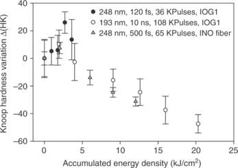
12.7.2 Fluoride glass fibers
While fluoride glasses were first realized in 1975 (Poulain et al., 1975), and shortly after their spectroscopic and active ion-host characteristics established them as promising materials for lasing devices (Zhu and Peyghambarian, 2010), their laser photosensitivity has been investigated in detail for almost two decades after the initial invention. The structure of fluoride glasses where the oxygen ion has been replaced by that of fluorine allows low phonon energies and thus great transparencies both at the Urbach tail band and the far-infrared regime, sketching an efficient bandwidth from 200 nm to 8 μm approximately. Poignant et al. recorded Bragg gratings in a ZBLAN fiber doped with Ce+3 ions as photosensitivity enhancer, reaching refractive index changes of few parts of ×10−4 after exposure to 246 nm pulsed radiation (Poignant et al., 1994; Taunay et al., 1994).
Since the photosensitivity observed was underlined by a specific sensitizer, potentially the mechanism of refractive index changes could be that of color-center formation; however, that was not concluded from the experiment (Taunay et al., 1994). Sramek et al. (2000) exposed fluorozirconate glass slabs of different compositions to 193 nm excimer laser radiation, and found that surface expansion occurs at heights of tens of nanometers for accumulated energy doses lower than 1 kJ/cm2 (Fig. 12.23). The last authors justified this laser-induced expansion after considering similar crystal structures such as b-BaZr2F10 and BaZrF6, which lead to density ratios close to unity after comparison with the fluorozirconate glasses exposed; thus exhibiting low structural defect population. The underlying photosensitivity mechanism speculated for 193 nm excitation, refers to Coulombian bond breaking between ion modifiers such as Ba with unpaired fluorine, changing the glass micro-coordination with respect to the Zr2F13 unit blocks.

12.23 Surface expansion of fluorozirconate glasses photo-induced at 193 nm as a function of the total energy impinging the glass (After Sramek et al., 2000.).
Moreover, Zeller et al. (2005) exposed undoped fluoride glasses to 193 nm excimer laser radiation and investigated the radiation-induced glass disorder by examining changes in the Urbach tail of the irradiated specimens (John et al., 1986; Urbach, 1953). Zeller and co-authors further quantified the depth of radiation damage into the exposed glasses by gradually polishing them and re-evaluating refractive index changes by means of Kramers–Kronig transformation of the color centers formed. Cerium doped ZBLAN fluoride bulks were also exposed by Williams et al. (1997) using 248 nm, excimer lasers, revealing that the Ce3+ excitation was of a two-step absorption nature, exploiting the 4f1–5d1 transition leading to quadratic dependence upon intensity. In the same study, it was also found that the Cerium ion color centers created were simultaneously bleached by the 248 nm excitation.
Similar to other Bragg grating inscription in low defect glass optical fibers Grobnic et al. (2006) used 800 nm femtosecond laser for recording ~ 2.0 × 10⁻⁴ refractive index changes in an undoped ZBLAN fiber; nonetheless, without referring to the sign of the refractive index changes. These gratings resisted to temperatures up to 240°C, quite close to the Tg of the glass at 265°C. An illustrative study on the photosensitivity of ZBLAN fibers was presented by Bernier et al. (2007), where negative refractive index changes were identified locally in the fiber core and cladding areas, using fiber profilometry (Fig. 12.24). Also, positive refractive index changes were traced at the core–cladding interface, and at other areas of the cladding, providing an initial evidence that the sign of the refractive index changes is directly dependent upon the dissipated intensity at microscopic level. Moreover, these refractive index engineering results appear to be in agreement with the volume dilation measurements in bulk samples presented by Sramek et al. (2000) and the Urbach tail investigations of Zeller et al. (2005) where a radiation-induced disordering of the glass is described.

12.24 Measured transmission and reflection spectra of a Bragg grating written in an undoped-ZBLAN LVF fiber at 0.9 mJ during a 2 s exposure using 800 nm femtosecond radiation. (After Bernier et al., 2007. Used with permission from Optical Society of America [OSA].)
12.8 Microstructured optical fiber (MOF) gratings
12.8.1 Bragg and long-period grating inscriptions
Photonic crystal fibers (PCFs) fuse unique light guiding properties together with useful microfluidic structures, offering great flexibility in developing photonic devices implemented with novel and high performance functionalities. Even though there was a standardized photosensitivity and refractive index engineering background related to the inscription of Bragg and long-period gratings in standard optical fibers and most of the glass materials used for drawing them, the emergence of PCFs and MOFs imposed new inscription issues as well as challenges (Knight et al., 1996; Russell, 2006). The capillary structure surrounding the guiding area affects the light penetration for side-illumination as this is principally required for periodic structure inscription using laser radiation. Specifically, the geometrically complex core and cladding shape of PCFs and MOFs induces significant scattering and refraction effects that in general reduce the average laser intensities delivered into the fiber core. Such reduced power dissipation into the fiber core during the side-illuminated grating inscription process deteriorates the photosensitivity yield by means of the magnitude of the refractive index changes formed into the guiding area and the accumulated energy densities needed for reaching saturation of the inscription. These scattering and refraction effects can dominate over the type of the photosensitivity process occurring, as well as the order of the underlying photon absorption. Many PCF and MOF designs are drawn from a single type of optical glass, often of low defect concentration such as fused silica, exhibiting low photosensitivity using conventional exposure methods. Therefore, additional issues arise during the grating recording in those high quality glass MOFs, rendering the inscription process a difficult and multiparameter materials science and light propagation problem.
Most of the methods used for inscribing gratings in standard optical fibers have been used for similar inscriptions in MOFs counterparts, revealing interesting grating recording behaviors and spectral responses. Initially, the safe path of Ge-based photosensitivity and hydrogenation was followed for forming Bragg and long-period gratings inside MOFs, by doping a small socket of the guiding area with Ge-ions.
Three years after the demonstration of PCF, Eggleton et al. (1999) presented the first Bragg and long-period grating inscription in such a solid core fiber (Fig. 12.25), which contained a Ge-doped photosensitive socket, using a 242 nm frequency-doubled excimer-pumped dye laser. For alleviating side-illumination scattering issues, Eggleton et al. hydrogen loaded the Ge-doped solid core PCF, reaching grating strengths greater than 50%. The same group presented a more detailed analysis of Bragg grating inscription and spectral resonance effects in several MOFs of different geometry and dopands, including excitation of cladding modes (Eggleton et al., 2000; Kerbage et al., 2000).

12.25 Measured transmission spectrum of a Bragg grating written in PCF (solid curve) before and (dashed curve) after application of the external index; the dotted curve shows the computer mode spectrum when beam-propagation modeling was used. (After Eggleton et al., 1999. Used with permission from Optical Society of America [OSA].)
Kakarantzas et al. (2002) presented the first long-period grating recording in a solid core all-silica PCF by using 10.6 μm CO2 laser radiation, and a scanning mirror beam focusing setup, following the technique inaugurated by Davis et al. (1998) in standard optical fibers. In this demonstration, the glass PCF was heated by the CO2 laser above the fictive temperature of silica, resulting in the formation of both geometrical and micro-coordination changes in the rapidly cooling glass and succeeding in inscribing short length, high strength LPGs and rocking filters (Kakarantzas et al., 2003).
Accordingly, the first Bragg grating inscription in an all-silica, solid core PCF was reported by Groothoff et al. (2003) using a 193 nm, excimer laser of standard spatial coherence and a phase mask in contact mode (Fig. 12.26). The Type I refractive index changes reported in that demonstration were of the order of 2.4 × 10⁻⁴, after an exposure of cumulative fluence of 200 kJ/cm2, leading to significant grating strengths. The refractive index modification mechanism was assumed to be that of two-photon resulting primarily in the dissociation of the Si-O bond, inducing matrix compaction. Such experimental observation is similar to the results presented by Rothschild (Rothschild et al., 1989), Devine (Fiori and Devine, 1986) and Borrelli (Borrelli et al., 1997). Also, these authors found that there were non-thermal, post-exposure matrix relaxation effects, resulting in spectral modifications of the inscribed reflectors. Canning et al. further exploited the above writing approach for developing DFB laser in an Er-doped silicate glass PCF, reaching slope efficiencies of 12.5% (Canning et al., 2003; Groothoff et al., 2005); but also in the recording of Bragg reflectors in a Fresnel type MOF (Martelli et al., 2005).

12.26 Grating wavelength evolution as functions of 193-nm cumulative fluence. (After Groothoff et al., 2003. Used with permission from Optical Society of America [OSA].)
Other groups used 193 nm excimer laser radiation for recording Bragg gratings in hydrogenated phosphosilicate and germanosilicate PCFs, reaching refractive index changes greater than 3 × 10⁻³ for the phosphosilicate matrix, and one order of magnitude higher changes for the germanosilicate fibers (Beugin et al., 2006). In the work of Beugin et al. (2006) the PCF section was spliced both sides to a standard optical fiber prior to hydrogenation, for encapsulating hydrogen under the loading pressure into the capillaries (Sørensen et al., 2005), maintaining high concentrations of hydrogen species into the PCF core during the grating recording process, while reducing the out-diffusion rate (Liou et al., 1997).
Violakis et al. (Violakis and Pissadakis, 2007b) used a high spatial coherence 193 nm excimer laser for recording strong Bragg reflectors in a commercially available (Fig. 12.27a), solid core PCF, reaching high refractive index changes (Δn = 3 × 10⁻⁴). The use of an increased spatial coherence laser allowed the formation of high contrast interference fringes along the whole cross-section of the exposed fiber (Othonos and Lee, 1995), a condition that is significant for maintaining high intensities necessary for nonlinear absorption photosensitivity processes. In the demonstration of Violakis et al. (Violakis and Pissadakis, 2007b) the Bragg reflectors recorded using an energy density of 230 mJ/cm2 exhibited a broad wide spectral valley observed at the left side of the fundamental grating notch, related with broadband coupling to radiation modes. This kind of radiation mode coupling was also confirmed when visible laser was injected into the PCF fiber and out-coupled by the inscribed Bragg grating. Such a spectral feature that is routinely observed in the case of surface relief gratings imprinted on planar waveguides (see Fig. 12.27b) may be related to a shallow, compaction relief periodic feature induced in the microstructured fiber capillaries (Schenker, 1994).

12.27 (a) Modulated refractive index versus accumulated energy density curves of Bragg grating inscriptions realized in the ESM-12–01, PCF, using energy density of 230, 170 and 90 mJ/cm2 using 193 nm, 10 ns laser radiation. (b) Transmission spectrum of a grating inscribed in the above fiber using 230 mJ/cm2 energy density per pulse and 187 kJ/cm2 accumulated energy density.
Fu et al. (2005) exposed a silica glass solid core PCF using a frequency tripled Ti:Sapphire 267 nm femtosecond laser for recording Bragg reflectors; however, the fiber was hydrogenated for increasing photosensitivity, similar to the example of Zagorulko et al. (2004). Using this approach the refractive index changes inscribed in the PCF exposed were ~ 3 × 10⁻⁴ under accumulated energy density reaching 100 kJ/cm2, while in a standard allsilica fiber three times greater index changes were recorded (1.1 × 10⁻³) using 54.5 kJ/cm2 (Zagorulko et al., 2004).
Similarly, Brambilla et al. (2006) used 264 nm, 220 fs laser radiation for inscribing 1 cm long and 20 dB strong LPGs into an all-silica solid core PCF. Different types of hydrogenated PCF fibers were also exposed to femtosecond and picosecond, 248 nm laser radiation using standard phase mask and double phase mask interferometer (Fig. 12.28), leading to refractive index changes of 1.2 × 10⁻⁴, for energy doses of ~ 18 kJ/cm2 (Pissadakis et al., 2008).
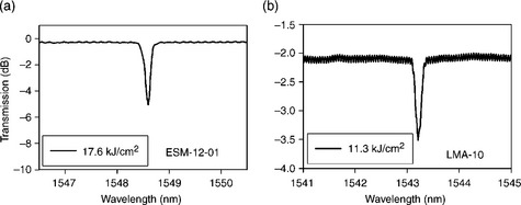
12.28 Transmission spectra of Bragg gratings fabricated in (a) ESM-12-01 and (b) LMA-10 hydrogenated optical fibers, having 1 cm length, by using 248 nm, 5 ps laser radiation. Accumulated energy densities appear in the spectra insets.
Other demonstrations included the use of Ti:Sapphire 800 nm, femtosecond laser radiation for Bragg grating inscription in pristine and tapered PCFs (Mihailov et al., 2006); and frequency tripped Ti:Sapphire laser at 262 nm for comparative recordings in unloaded and hydrogen-loaded PCFs (Wang et al., 2009b). All of the above demonstrations led to the formation of Type I photosensitivity refractive index changes and inscription behavior; however, there were examples where Type IIA Bragg reflectors were inscribed in MOF and PCF using 193 nm excimer lasers. Cook et al. (2008) exposed a 6.7%wt Ge-doped core, 12-ring PCF for completing Type IIA saturation after typical accumulated doses of 20 kJ/cm2. Others (Pissadakis et al., 2009b) exposed a 36%wt Ge-doped, small-core, highly nonlinear PCF, for observing complex Type IIA photosensitivity effects for the two guiding modes supported by the fiber (Fig. 12.29a). Both scattering modes exhibited post-fabrication, amplification effects after storing at room temperature due to stress relaxation of the small dimension 1.38 μm, highly doped core; while surviving up to 800°C following a mixed Type I and IIA decay behavior. (see Fig. 12.29b)
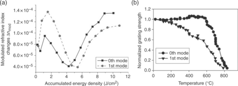
12.29 (a) Index modulation Δnmod changes versus accumulated energy density, monitored for the 0th and 1st order guiding modes of the MOF, for Type IIA Bragg grating exposure using 193 nm, 10 ns excimer laser radiation. (b) Isochronal annealing results for Type IIA Bragg gratings recorded in high-Ge microstructured fiber, using 193 nm excimer laser radiation. Exposure conditions for the ESM-12–01 fiber: 193 nm, 230 mJ/cm2 energy density, 200 kJ/cm2 accumulated energy dose. The ESM-12–01 fiber has been annealed at shorter isothermal intervals of 10 min.
In parallel, there is interest in the inscription of Bragg reflectors in small and collapsed core microstructured optical fibers. Due to the specific core geometry and dimensions (< 5 μm), collapsed core fibers exhibit a guiding mode profile largely extending in the inter-capillary areas, allowing high overlap with the refractive index medium included in those. The last renders them ideal platforms for the development of high sensitivity in-fiber refractometers and biological sensors, prompting the inscription of Bragg reflectors in their minimal size cores.
Initially, there were reports on the Bragg grating inscription in hydrogenated, Ge-socket collapsed core microstructured optical fibers using CW, 244 nm, frequency doubled Ar+ ion laser radiation (Huy et al., 2006; Phan Huy et al., 2007) (see Fig. 12.31b, c). Becker et al. (2009) used 267 nm, femtosecond laser radiation for recording arrays of Bragg reflectors in all-silica collapsed core fibers (Fig. 12.30b,c), reaching refractive index changes of 6 × 10⁻⁴.

12.30 (a) Transmission spectrum of FBG photo-written in the three-hole fiber over a spectral window of 50 nm, showing spectral resonances toward high-order modes. The inset shows a zoom on the Bragg wavelength itself. (After Phan Huy et al., 2007. Used with permission from Optical Society of America [OSA].) (b) Target fiber IPHT-256b1 with grating arrays. Microscope picture. The black bar is the scale for 50 μm. (c) Reflection spectrum of a fiber Bragg grating array with four gratings. (After Becker et al., 2009. Used with permission from IEEE.)
Recently, Ge-doped socket, collapsed core fibers were exposed using high coherence, 193 nm, excimer laser radiation (Fig. 12.31), leading to visible compaction effects in the exposed core, resulting in significant light out-scattering for shorter wavelengths (Konstantaki et al., 2011). The structural changes induced in the collapsed core and surrounding cladding are under further investigation using micro-Raman analysis.

12.31 (a) Reflection spectrum of a Bragg reflector inscribed into a Hi-Ge socket, collapsed core fiber using phase mask and 193 nm, 10 ns excimer laser. Exposure conditions: 150 mJ/cm2, 24 K pulses. (b) Optical microscope photo of the collapsed fiber core after the above grating inscription.
Novel Bragg grating inscription approaches, while exploiting the absorption or transparency of the infiltrated/encapsulated material into the PCF capillaries, have been presented for all-solid PCFs. Jin et al. (2007) attempted Bragg grating inscriptions using 248 nm excimer laser, in a hydrogenated, all-solid silica PCF containing into the core surrounding capillaries 1%wt Ge-doped silica glass, reaching ~ 1 dB reflectivities, and cladding mode inversion at the red-side of the spectrum. In another fashion, Bigot et al. (2009) designed an all-solid PCF, where the capillaries contained a low-photosensitivity, phosphosilicate glass, while the grating host material was a F/Ge co-doped core. In such an arrangement, the low photosensitivity glass infused into the capillaries did not impose absorption issues for side irradiation at the recording wavelength, allowing significant intensities reaching the fiber core and thus inducing refractive index modulation.
12.8.2 Studies on the issue of the side-illumination scattering during grating inscription
In all of the above Bragg and long-period grating demonstrations utilizing ultraviolet laser sources, there was not any special preparation related to the relative rotational position of the fibers with respect to the side-illuminating laser beam, therefore, the photosensitivity and refractive index engineering results presented there were not absolutely accurate or repeatable. The first study on the impact of the capillary structure and its rotational or transversal position along the fiber axis with respect to the laser beam on the Bragg grating recording processing was presented by Marshall et al. (2007).
Experimentally, Marshall et al. utilized 267 nm, femtosecond radiation coupled under different conditions into the PCF under exposure for different relative orientations and positions, and accordingly the 1.9 eV photoluminescence (Stathis and Kastner, 1984) induced was measured through the PCF fiber core. The same group used a finite element method for simulating the propagation of a small size beam for the aforementioned side-illuminating conditions evaluating an average field intensity (Fig. 12.32), and correlating such measurements with those obtained from photoluminescence experiments for different angles of rotation (Marshall et al., 2007). Similar investigations were presented by Canning et al. (2008a) using finite difference time domain (FDTD) simulations for estimating the intensities reaching the PCF core for a variety of rotational angles during Bragg grating side-illumination, for 193 nm excimer laser radiation. Geernaert et al. (2008) inscribed Bragg gratings in a highly asymmetric germanium-doped, birefringent microstructured optical fiber using a 248 nm excimer laser that had been subjected to hydrogenation; thus, strong single-photon absorption effects dominated its photosensitivity. Geernaert et al. found that the rotational state of the fiber around its axis during this single-photon absorption inscription did not significantly affect the reflection strength of the Bragg grating, while having a greater impact in its wavelength.
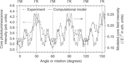
12.32 TE polarization PCF core photoluminescence intensity and computational model wholecore field intensity as a function of PCF rotation angle for the wavelength of 267 nm. (After Marshall et al., 2007. Used with permission from Optical Society of America [OSA].)
In another approach the FDTD method was used again for illustrating in greater detail the average, maximum and minimum intensities reaching the core of all-silica standard and microstructured optical fibers (Fig. 12.33), leading to better insights with respect to the impact to the material photosensitivity (Pissadakis et al., 2009a). In that study, the wavelength investigated was that of 248 nm, for 5 ps pulse duration and for propagation into hydrogenated silica glass fibers. The simulation results obtained depicted that while the mean intensities occurring in a side-illuminated PCF were 30%–40% lower than those found in a standard all-silica fiber, the maximum intensities that can be found in the core of a side-illuminated PCF can even be 200% greater (Fig. 12.34).
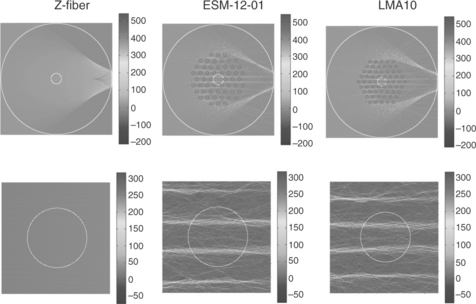
12.33 Simulation results of the spatial distribution of the Pointing vector S using the FDTD method for the cases of Z-fiber, ESM-12–01 and LMA10 for 0 ° rotation angle. The outer and inner white circles define the fiber cladding and core, respectively. The grey-scale bar corresponds to the scale of the values of the Poynting vector S measured in GW/cm2. The dark circles define the spatial boundaries of the individual capillaries. (Upper row) Simulation of the side-illumination corresponding to views of the whole fiber areas with dimensions 126 μm × 126 μm. (Lower row) Simulation of the side-illumination corresponding to the vicinity of the fiber core of the above optical fibers, for core diameters 9 μm (Z-fiber), 12 μm (ESM-12–01) and 10 μm (LMA10).
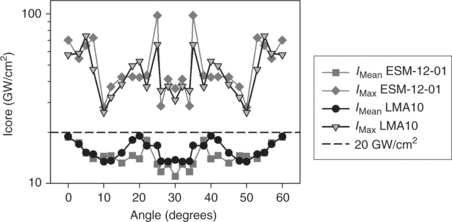
12.34 Intensity figure in the core area Icore versus rotational angle with respect to the ΓK axis for the ESM-12–01 and LMA10 microstructured optical fibers. IMean: Averaged intensity. IMax: Maximum intensity. The IMean values have been evaluated for the circular areas presented in Fig. 12.33. The IMax figure has been sampled for a square patch of 0.2 μm side, around the maximum points of intensity. The dash line refers to input pulse intensity as that measured before reaching the fiber complex.
Scattering can greatly affect the triggering of specific photosensitivity processes, especially those of two-photon absorption which are quadratically dependent upon the local intensities, and dominate the refractive index engineering. In this study, the standard and PCF all-silica fibers exposed reconfirmed that the Bragg gratings recorded in the PCF were of lower strength than those formed in the standard all-silica fiber (Fig. 12.35).
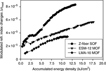
12.35 Modulated Δnmod refractive index changes versus accumulated energy density for grating recordings in the standard optical fiber (Z-fiber), and in the PCFs, ESM-12–01 and LMA10. The refractive index data have been normalized to the corresponding mode confinement factor of each fiber.
The issue of laser beam scattering for side-illumination during Bragg grating recording has been also addressed by infiltrating inside the fiber capillaries liquids of high transparency at the irradiation wavelength and of refractive index value close to that of silica; or by suitably designing the fiber capillaries’ geometry for scattering less. This approach can lead to improved recording results, in the case where the fiber core is highly photosensitive, with linear absorption coefficient substantially higher than that of the infiltrated liquid. Sørensen et al. (2007) infiltrated organic solvents into a hydrogenated, Ge-doped core PCF for reducing the deteriorating effect of extensive side-illuminating scattering during Bragg grating recording using 248 nm pulsed laser radiation. Methanol and n-heptane reduced scattering effects which allowed significant amount of photons to reach the Ge-doped PCF core and induce significant refractive index changes, improving the grating strengths obtained by several decibels.
Following a different route, Baghdasaryan et al. (2011) used the FDTD method for simulating different candidate geometries with respect to the d/λ factor and hole diameter for reducing side-illumination scattering for 800 nm and a specific pulse duration. The results obtained in Baghdasaryan et al. (2011) provide a first indication that such optimization is possible without sacrificing the guiding properties of the custom-made PCF. Petrovic, Allsop, et al. (Allsop et al., 2011; Petrovic and Allsop, 2010) examined the effect of the numerical aperture of the focusing optics used during grating inscription in PCF using femtosecond lasers, further studying the effect of the specific recording conditions in the annealing and spectral characteristics of the inscribed gratings, thus providing a correlation with the photosensitivity mechanisms triggered.
12.9 Laser machining of optical fibers
The modification and structuring of optical materials surfaces have been constantly progressed since the early years after the development of the laser sources; greatly benefiting from the emergence of high power lasers (Bäuerle, 2000). While progress in the laser surface structuring and ablation field has continued since the early 1980s for modifying the surface of planar samples (Endert et al., 1995), the structuring of optical fibers progressed at a later time. The last is mainly attributed to three parameters: (i) optical fibers are species of small physical dimensions, needing precise translation and beam focusing systems for achieving machining over their limited end-face or curved cladding; (ii) optical fibers are mostly drawn from the highly transparent silicate glasses exhibiting poor surface etching quality using most of the nanosecond laser sources available at that period (Ihlemann et al., 1992); (iii) the potential and advantages of optical fiber surface micro-structuring were not illustrated at that time. Therefore, the examples of such microstructuring were quite few, mainly focused on the melting of micro lenses onto fiber end-faces (Paek and Weaver, 1975; Presby et al., 1990) using CO2 lasers and the drilling of via holes into optical fibers or fiber-like structures (Gower, 2000, 2001).
The state-of-the-art in the optical fiber machining field was enriched after high spatial accuracy, ultraviolet nanosecond (mainly 157 nm excimer laser) and infrared femtosecond laser micromachining systems became available (Gattass and Mazur, 2008; Vainos et al., 1996), in conjunction with the demonstration of promising results in the high quality etching of highly transparent optical materials (Dyer et al., 2003; Jackson et al., 1995; Jürgen et al., 2007). Together, the emergence of PCFs (Knight et al., 1996) and the lab-on-chip biosensing and microfluidic architectures (Haeberle and Zengerle, 2007; Oosterbroek and Berg, 2003) further underlined the advantages and possible functionalities that a laser-machined optical fiber can offer in developing advanced photonic (Monat et al., 2007) and optofluidic (Psaltis et al., 2006) systems. The cylindrical geometry of optical fibers and the small area of the fiber end-face frustrate the use of standard photolithographic techniques to be applied in structuring them. In addition, the use of laser beams for such surface modification offers distinct advantages compared to other approaches: via hole opening and drilling can be driven along the whole diameter of the fiber at reasonable etching times, while rotational machining such as lathing can be easily performed; real-time in-fiber spectral monitoring of the etching is also possible.
The favorable lasers for silicate glass optical fiber machining are these of 157 nm, excimer nanosecond and 800 nm, Ti:Sapphire femtosecond lasers. The 157 nm excimer laser radiation offers single-photon absorption interaction for the silicate glasses irrespective of dopands and impurities concentration, while etching rates achieved are of the order of a few tens of nanometers per pulse, solely dominated by volume absorption within shallow depths (Dyer et al., 2003; Jürgen et al., 2007; Li et al., 2007). Etching quality is of highest quality for wide area pits even after several pulse exposures, where incubation effects are in favor of the etching quality. On the other hand, ablation of silicate glasses using 800 nm, or other near-infrared femtosecond lasers are dominated by multiphoton absorption triggering avalanche ionization and plasma formation effects over minimal volumes defined by the intensity pattern of the focused beam (Liu et al., 1997; Sugioka et al., 2005). Etching rates are generally greater than those obtained using ultraviolet sources, allowing machining in shorter time intervals of deeper trenches. The etching quality of the structures is highly dependent upon the specific parameters of the exposure, where heat accumulation effects (Eaton et al., 2005; Schaffer et al., 2003) can substantially affect the surface quality obtained. In general, surface or volume structuring using near-infrared femtosecond sources is accompanied by post-fabrication chemical conditioning or etching process for achieving improved roughness of the exposed areas or for etching deep channels into the materials volume (He et al., 2010). Moreover, there are two main approaches in the laser machining of optical fibers: that of end-face and that of cladding surface/volume structuring. Examples of the two above machining fashions will be reviewed below, describing the etching conditions and structure design and the photonic functionality imparted into the etched optical fiber; the new approach of ‘in-fiber’ structuring will be also presented.
12.9.1 End-face machining
The most frequent structures formed onto the cleaved end-face of standard optical fibers were these of diffraction gratings, micro lenses, interferometers and Fabry-Perot cavities. These optical structures combine relative straightforward etching processing protocol, while providing significant photonic operations such as tailored guiding mode out-/in-coupling, spectral analysis and optical phase reading. The simplest micromachining approach followed by several groups was that of local heating of cleaved fiber ends using 10.6 μm CO2 laser beams, for forming end-firing, convex micro lenses (Abdelrafik et al., 2001; Presby et al., 1990); however, this achieved rather low control over the shape of the lenses formed (Fig. 12.36a). Li et al. (2007) used a 157 nm excimer laser for etching diffraction gratings of 2 μm period, micro lenses and Mach–Zehnder step-shifted interferometer onto the endface of multimode and standard telecom fibers. In this work energy densities greater than 4 J/cm2 were employed for etching smooth pits and features onto the cleaved silicate glass fiber facet, reaching 5 μm flat depth pits, after single-photon absorption interaction.
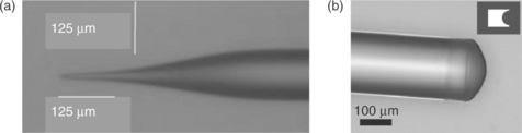
12.36 (a) Optical micro lens fabricated in an optical fiber using CO2, 10.6 mm laser radiation, following a two-step process. (After Abdelrafik et al., 2001.) (b) An optical microscopy image of a micro lens-tipped optical fiber using 157 nm, excimer laser radiation (After Li et al., 2007.).
Parabolic end-face lenses were formed using lathing machining schemes (Fig. 12.36b) while utilizing Schwarzschild reflecting objective and 157 nm radiation (Dou et al., 2008). Excimer laser 157 nm radiation was also employed by Machavaram et al. (Machavaram, 2006) for end-face pit and sharp tip machining in several fiber types (including sapphire fibers), in combination with selective chemical etching post-processing for improving surface smoothness and achieve specific shaping. The same wavelength was used by Ran et al. (2009) for initially forming flat-top, high smoothness, ~ 30 μm deep pits on SMF-28 optical fiber, and then enclosing these etched cavities into the fiber line by fusion splicing, forming high finesse Fabry-Perot interferometers. This in-fiber Fabry-Perot etalon exhibited spectral fringe contrast of 30 dB, allowing strain measurements at high temperatures and modulation rates. Using a more complex splicing, cleaving and re-ablating process an end-face refractometer was presented by the above group providing resolution of 1130.887 nm/RI units (Zengling et al., 2009).
Using different laser wavelength (785 nm) and pulse duration (125 fs) than nanosecond 157 nm excimer laser radiation, 33° angle (see Fig. 12.37), conical shape pits were formed into optical fibers for side-firing out-coupling (Sohn et al., 2010). The relatively rough ablated surface was polished to better quality using post-exposure arc discharging. Alternatively, by adapting a suitable length splicing buffer fiber to a PCF, and subsequent machining holes through the end-face of this buffer layer, Wang et al. (2010) presented a selective infiltration technique of the surrounding micro-capillary structure.

12.37 Optical microscope image of the optical fiber tip microstructured by using a femtosecond laser. (After Sohn et al., 2010. Used with permission from Optical Society of America [OSA].)
12.9.2 Structuring through the cladding
The structuring of the fiber end-face using laser beams led to the development of optical fiber structures exhibiting purely optical functionalities. The structuring through the cladding area of optical fibers using laser processes allowed the implementation of microfluidic functionalities in addition to the photonic structures developed until then. For a long period, the related state-of-the-art was rather primitive where simple etched holes (Gower, 2000) and jacket removal (Barnier et al., 2000) only had been presented. Then the use of 157 nm and 800 nm-femtosecond laser micromachining systems prompted the versatile structuring of small size photonic devices such as optical fibers.
Deeper through-out trenches, of several tens of micrometers’ length, were ablated in SMF-28 optical fibers using 157 nm excimer laser radiation by several authors for forming rectangular shape air cavities (Fig. 12.38a) and observing Fabry-Perot interferometric spectral modulations (Fig. 12.38b) in transmission and reflection mode (Machavaram et al., 2007; Ran et al., 2007, 2008); others used 800 nm, 1 KHz femtosecond radiation for ablating similar long length cavities (Li et al., 2008). Such demonstrations were also reported for in-fiber etalon etching in endlessly single mode PCFs allowing fringe probing for wider wavelength bandwidths (Ran et al., 2007; Rao et al., 2007). In most of these Fabry-Perot in-fiber interferometers a primary concern was related to the flatness and parallelism of the opposite walls constituting the oscillating cavities along the longitudinal axis of the fiber.

12.38 SEM picture of an ablated cavity through the diameter of the optical fiber at 25 × 104 J/m2. Fabry–Perot interference fringes produced from cavities micromachined at an energy density of 25 × 104 J/m2 in SMF-28 with 47.5 μm cavity length (After Machavaram et al., 2007.).
Zhou et al. (2007) used 800 nm femtosecond laser volume damage micro-machnining, in combination with HF acid selective chemical etching for forming narrow slot openings and boreholes in the cladding of optical fibers, suitably positioned within the length of pre-inscribed photosensitive Bragg gratings. These inscribed slots allowed the infiltration of these microfluidic fiber Bragg gratings using fluids with different refractive indexes, developing refractometers of 945 nm/RIU sensitivity. A similar micromachined hole design in a standard optical fiber was used for refractometric measurements where the light was refracted and out-coupled from the cylindrical shape ring upon its refractive index contrast with the fiber core (Wang et al., 2009a). Selective chemical etching of femtosecond laser-exposed Bragg gratings using a phase mask was used for forming deep periodic structures into a standard optical fiber (Fig. 12.39) and realizing infiltrated refractometer by monitoring the shift and power change of the Bragg peak scattered (Yang et al., 2011).

12.39 Microscope images of the fs laser-induced FBG before and after chemical etching. (a) Side view normal to the beam axis and (b) crosssection before etching. (c) Side view normal to the beam axis and (d) view from output side of laser beam after etching. (e) SEM image on the fiber grating surface after etching. (After Yang et al., 2011. Used with permission from Optical Society of America [OSA].)
The laser etching of the cladding of microstructured and photonic crystal fibers provided a novel approach in the optimum exploitation of the micro-/optofluidic properties of these fibers. A first example was presented by van Braken et al. (2007), using an 800 nm femtosecond laser for drilling microchannels reaching the photonic bandgap periodic structure of a hollow core PCF; also in the same work micro-channels were drilled in collapsed core fiber claddings. Such micro-channels were exploited for side pressurized C2H2 gas infiltration and optical absorption measurements at the 1.5 μm band. Trench etching of the fiber cladding using femtosecond infrared laser radiation was used as an alternative method for selectively filling of the capillaries of a solid core PCF, by removing an arc slice of the out-cladding and reaching capillary channels lying underneath, exposing them for liquid immersion (Yiping et al., 2010). Controlled hole drilling with respect to the depth toward the fiber core was achieved using 800 nm femtosecond radiation, for developing long-period rejection filters in solid core all-silica PCF (Liu et al., 2010).
CO2 lasers emitting at 10.6 μm do not provide structuring of high spatial resolution and most applications were presented on thermal lensing formation onto fiber end-faces. Nonetheless, after the demonstrations of Davis et al. (1998) and Kakarantzas et al. (2002), there several other examples were reported that laser radiation could be used for fine cladding structuring of standard and microstructured fibers. Wang et al. (2006) used CO2 laser radiation for etching grooves onto standard optical fiber claddings for forming strongly scattering LPGs, while later presenting the thermal cladding deformation in hollow core PCFs demonstrate the first LPG in such a fiber design (Wang et al., 2008). Another group used a CO2 laser beam in a lathing setup for inducing periodic, helical deformations outside of a standard optical fiber for realizing torque sensitive band rejection filters (Oh et al., 2004).
12.9.3 ‘In-fiber’ structuring using lasers
The use of laser radiation for modifying/etching the inner walls/capillaries of PCFs and MOFs has not been broadly investigated yet. In such an approach the laser beam passes through the cladding area and interacts selectively with the channels enclosed; the fiber core can be also modified. The main problems related to the in-fiber etching approach are those of extensive damage of the structure due to the explosive nature of ablation, deteriorating transmission signal and increasing losses; as well as the difficulty in controlling the exposure conditions and gas infiltration pressure for maintaining etching near the fiber core. Shujing et al. (2010) exploit the debris products of in-fiber ablation generated by 800 nm, femtosecond laser beam for selectively filling the capillaries of a solid core PCF and form a kind of damaged long-period gratings in those fibers; while achieving diffraction efficiencies of more than 20 dB over few (< 15) grating periods.
Violakis et al. (Violakis and Pissadakis, 2007a) attempted for the first time the etching of Bragg gratings into the capillaries of solid core PCFs infiltrated with fluorinated gas, using 193 nm excimer laser radiation. For simple gas infiltration inside the fiber capillaries under atmospheric pressure, some exposure results indicated that the existence of SF6 inside the fiber capillaries can promote relief structure generation. While SF6 exhibits high selectivity over silica glass etching, attacking preferentially silicon (Chuang, 1981), its photo-dissociation inside the PCF channels could offer controlled etching of the capillary walls (Batool et al., 2004). However, the method did not lead to easily reproducible results, due to extensive redeposition of ablated glass and reaction products inside the capillaries (Batool et al., 2004). Also, the uncontrolled SF6 gas pressure conditions, where no buffer gas was used, while maintaining high pressures, increased recombination rates of excited species of fluorine.
Gratings fabricated in SF6 infiltrated PCFs, exhibited increased strengths and faster saturation under specific exposure conditions by means of energy density and number of pulses (Fig. 12.40). Nonetheless, SEM pictures of cleaved fibers at the grating exposed section, revealed extensive debris redeposition and hole blocking. The last can be responsible for the saturation of the Bragg grating etching process earlier than inscriptions performed in empty PCF using identical conditions.

12.40 Bragg grating recording in empty and SF6 gas infiltrated ESM-12–01 optical fiber using 193 nm, 10 ns excimer laser radiation and a phase mask.
Recently, Sozzi et al. (forthcoming) have employed fluorinated organic liquids infiltrated inside MOFs and PCFs for etching Bragg and long-period gratings into their capillary structure using 248 nm, 5 ps laser radiation. By suitably tailoring the absorption coefficient of the infiltrated medium with respect to the capillary structure and geometry, poor etching and non-repeatability issues reported by Violakis et al. can be minimized, allowing efficient in-fiber periodic structuring (Fig. 12.41).

12.41 SEM pictures from relief Bragg gratings etched inside the capillaries of a grapefruit optical fiber, using 248 nm, 5 ps excimer laser radiation and fluorinated gas infiltration. (a) Far view of the etched fiber cross-section. (b) Zoomed view in a single capillary area. The MOF fiber used was provided by ACREO. The fiber has not been washed after etching process for detaching from the machined area debris deposition.
12.10 Future trends and prospects
The methods, results and developments presented above, primarily related to optical fiber photosensitivity and the mirror-field of laser structuring, reveal immense progress with respect to the founding experiments of Weeks, Primak and Hill, reported more than three decades ago. This great but unpredictable progress was fertilized by three basic parameters: the continuous development of new and efficient laser sources and processing methods, the deeper understanding of the underlying laser–matter interactions, and the evolution of the optical fiber from a simple optical cable to a versatile and efficient photonic device platform. While the two former aforementioned parameters catalyzed the material science of laser photosensitivity and structuring, the last prompted the innovation effort for integrating the results emerging from fundamental and applied studies into the development of real photonic devices.
Reviewing the results over the last three decades, one may conclude that the optical fiber photosensitivity field has reached maturity mainly by solving two cross-related problems: the inscription of large and controllable refractive index changes into almost any fiber available, irrespective of material, dopand concentration and pre-conditioning, by using fast and reasonably priced techniques, suitable for mass production applications. The last statement is quite accurate and describes well the case of standard optical fibers, where the laser methods and yields for the inscription of Bragg and long-period gratings have fulfilled even the most demanding industrial or academic expectation. The use of high photon energy (ultraviolet) and/or ultrafast pulse duration (femtosecond and picosecond) laser sources can bridge the bandgap of most optical materials by exploiting specific defect annihilation or multiphoton absorption and correlated effects, allowing inscription of exploitable refractive index changes. Notably, the fact that the laser sources needed for performing such material modifications are commercially available providing high intensity and energy density outputs, at relatively modest costs, prompts the broader exploitation and improvement of the photosensitivity approaches investigated.
Alternative methods leading to the inscription of refractive index changes into high bandgap optical materials, exhibiting specific optical, annealing, radiation resistance or mechanical properties can be issues for future investigation. These methods may include the use of more than two laser sources irradiating the fiber sample simultaneously or with specific pulse delays, for triggering or suppressing photosensitivity mechanisms and thus controlling the type and properties of refractive index changes inscribed (Obata et al., 2002). Another approach of further interest is that of filamentation, for inscribing Bragg or long-period gratings in undoped silicate or other soft glass fibers, boosting diffraction efficiency and thermal stability of the reflectors (Bernier et al., 2011). The topic of thermally regenerated and photochemically induced gratings can also be further investigated/improved with respect to the amplification characteristics of the gratings and the underlying physiochemical mechanisms involved (Fokine, 2002a; Lindner et al., 2011). Such regenerated gratings may serve as durable radiation sensors (Gusarov et al., 2007) operating in harsh environments. Another route toward the improvement of the thermal stability and radiation behavior of fiber grating structures can be that of the adoption of alternatives to Ge ion dopands for modifying the material properties of the glasses used for fiber drawing, and accordingly the type of defects and glass transformation properties of the fibers (Butov et al., 2006; Shen et al., 2004).
Even though the grating inscription processing in standard optical fibers is based on well-established techniques and the photosensitivity mechanisms are understood to a great extent, there is a lot of room for new investigations and further improvements related to the grating inscription in microstructured and PCFs using laser radiation. The main issue arising from the capillary structure surrounding the solid or hollow guiding core is that of beam scattering for side-illuminations during grating recording. This important issue has been addressed by several groups while employing a number of simulation methods and experimental techniques (Canning et al., 2008a; Geernaert et al., 2008; Marshall et al., 2007; Pissadakis et al., 2009a). All these studies have agreed as to the deleterious role of the capillary structure constituting the PCFs and MOFs; however, they have not yet addressed the exact growth of the induced refractive index changes for different photosensitivity models (single- or multiphoton), and the overlap of these index changes with the guiding modes in the fiber core. Moreover, recently there has been effort made in the optimization of the Bragg grating recording conditions, either by liquid filling the fiber capillaries, or by designing a fiber capillary structure that results in reduced scattering during side-illumination, without significantly affecting the guiding properties (Baghdasaryan et al., 2011). The optimization of the combination between the PCF/MOF geometry and the recording wavelength and pulse duration for maintaining high photon fluencies reaching the fiber core is a hot topic that will address a challenging technical problem with direct practical impact. Such studies may also include the design of PCF/MOF with tailored spatial characteristics, dopand concentration within the fiber core or cladding for maximizing the overlap with the side-illuminating beam, for alleviating inscription complexity and repeatability obstacles. The infiltration of highly photosensitive polymeric matrices inside all-silica glass PCFs, and the recording of Bragg gratings in those infiltrated capillaries, and not into the highly transparent glass, constitutes a promising approach which can be directly exploited in the future for rendering the grating inscription in such fibers a rather easier task (Kakarantzas et al., 2011).
The structuring of optical fibers using deep, ultraviolet and infrared femtosecond lasers exhibits greater prospects with respect to novel future developments. Among others, there are two future laser machining directions that may lead to the development of novel optical fiber devices for sensing and actuating applications. First, the laser micro-/nanostructuring of standard and microstructured optical fibers, for generating complex and functional fluidic and diffracting elements in the cladding and core areas of those. Second, the cladding of standard but mostly of microstructured optical fibers can constitute a ‘free-space playground’ where the laser etching techniques described above can be used for inscribing relief or perforated structures and channels which can either allow controlled liquid/gas infiltration, fluid-trapping or localized mode perturbation. Such photostructuring engineering, together with photosensitivity changes inscribed and other selective chemical etching methods, will allow the realization of complex and versatile optofluidic circuits, which can constitute the ‘lab-in-fiber’ approach, following the example of the ‘lab-on-chip’. Such ‘lab-in-fiber’ photonic devices can find sensing applications in biology, instrumentation or medicine, implementing more than one functionality into a single photonic element.
Other advanced laser processing approaches can include new etching techniques such as laser-induced backside wet etching (Böhme et al., 2002) for structuring the inner walls of MOFs and PCFs, without affecting the out-cladding area, by infiltrating the fiber capillaries using highly absorbing liquids at the wavelength of the exposure. Other similar material sputtering techniques that can be applied in the case of MOFs and PCFs are those of laser chemical vapour deposition (LCVD) and laser-induced forward transfer (LIFT) for creating relief hetero-material structures inside their capillaries (Arnold and Pique, 2007; Klini et al., 2008).
Non-ablative methods are described in the use of femtosecond laser for inscribing relief (Do Lim et al., 2009) or photo-polymerizing organic–inorganic relief optical elements onto the end-face of optical fibers may also attract academic and industrial interest. Such ‘on-fiber’ hybrid structures can be photo-polymerized by self-guiding processes (Soppera et al., 2009) or by utilizing external femtosecond laser beams for creating a variety of guiding, refracting and diffractive elements by multiphoton absorption processing, at sub-wavelength resolutions (Malinauskas et al., 2011; Williams et al., 2011a). By applying specific chemical affinity processes onto these organic–inorganic structures, new kinds of chemical and biological sensing probes can be realized, exhibiting the advantages and functionalities of planar geometries into the cylindrical optical fiber topology.
12.11 Conclusions
The use of laser radiation for photosensitization, refractive index engineering and structuring of optical fibers has been one of the fastest growing topics in photonic science and technology during the last three decades. The majority of the scientific background and technological innovation accumulated during this period has found several commercialization paths in the service of everyday applications ranging from telecommunications and sensing to health and metrology. The refractive index engineering of glass optical fiber that dominated the field has reached maturity, leading to an increase of the photo-induced refractive index yields by more than four orders of magnitude, reaching levels of ×10−2 or greater. Simultaneously, new technological challenges are opening in the mirror-field of surface micro-/nanostructuring and ablation of standard and microstructured optical fibers, using high intensity and photon energy laser beams, realizing and embedding microfluidic operations into the fiber geometry. The critical idea is to judiciously combine and complement the fields of laser processing, photosensitivity and surface structuring in order to bring a powerful fabrication tool into the realization of future fiber optic and photonic devices.
12.12 Acknowledgments
The author would like to acknowledge the contribution of his colleagues Maria Konstantaki, Georgios Tsibidis, Paul Childs and Aashia Rahman for their scientific work in the fields of fiber photosensitivity and optical fiber structuring. Moreover, the author would like to thank the vast work of the talented PhD and MSc students Christos Pappas, Georgios Violakis, Michalis Livitziis, Irene Michelakaki and Michele Sozzi for their valuable findings during their dissertation studies. Several parts of the work presented above have been funded by research projects of the European Commission, while fiber and glass samples have been kindly provided by Sumitomo Europe, Schott USA, IPHT-Jena, Fibercore Ltd and ACREO SA. Finally, the author would like to thank the Optical Society of America (OSA) and the Institution of Engineering and Technology (IET) for kindly providing copyrighted material included in this chapter.
12.13 References
Aashia, R., Madhav, K.V., Ramamurty, U., Asokan, S. Nanoindentation study on germania-doped silica glass preforms: Evidence for the compaction-densification model of photosensitivity. Optics Letters. 2009; 34:2414–2416.
Abdelrafik, M., Renaud, B., Frederic Van, L. Two-step process for micro-lens-fibre fabrication using a continuous CO2 laser source. Journal of Optics A: Pure and Applied Optics. 2001; 3:291.
Albert, J., Fokine, M., Margulis, W. Grating formation in pure silica-core fibers. Optics. Letters. 2002; 27:809–811.
Albert, J., Hill, K.O., Johnson, D.C., Bilodeau, F., Mihailov, S.J., Borrelli, N.F., Amin, J. Bragg gratings in defect-free germanium-doped optical fibers. Optics Letters. 1999; 24:1266–1268.
Albert, J., Malo, B., Hill, K.O., Bilodeau, F., Johnson, D.C., Theriault, S. Comparison of one-photon and 2-photon effects in the photosensitivity of germanium-doped silica optical fibers exposed to intense arf excimer-laser pulses. Applied Physics Letters. 1995; 67:3529–3531.
Albert, J., Schulzgen, A., Temyanko, V.L., Honkanen, S., Peyghambarian, N. Strong Bragg gratings in phosphate glass single mode fiber. Applied Physics Letters. 2006; 89:101127.
Allsop, T., Kalii, K., Zhou, K., Smith, G.N., Komodromos, M., Petrovic, J., Webb, D.J., Bennion, I. Spectral characteristics and thermal evolution of long-period gratings in photonic crystal fibers fabricated with a near-IR radiation femtosecond laser using point-by-point inscription. Journal of the Optical Society of America B. 2011; 28:2105–2114.
Archambault, J.-L.Photorefractive gratings in optical fibres. University of Southampton, 1994. [[Online]].
Archambault, J.-L., Reekie, L., Russell, P.S.J. 100% reflectivity Bragg reflectors produced in optical fibres by single excimer laser pulses. Electronics Letters. 1993; 29:453–455.
Armitage, J.R. Fiber bragg reflectors written at 262nm using a frequency quadrupled diode-pumped Nd3+-YLF laser. Electronics Letters. 1993; 29:1181–1183.
Arnold, C.B., Pique, A. Laser direct-write processing. MRS Bulletin. 2007; 32:9–12.
Atkins, R.M., Mizrahi, V., Erdogan, T. 248 nm induced vacuum UV spectral changes in optical-fiber preform cores: Support for a color center model of photosensitivity. Electronics Letters. 1993; 29:385–387.
Baghdasaryan, T., Geernaert, T., Berghmans, F., Thienpont, H. Geometrical study of a hexagonal lattice photonic crystal fiber for efficient femtosecond laser grating inscription. Optics Express. 2011; 19:7705–7716.
Bandyopadhyay, S., Canning, J., Stevenson, M., Cook, K. Ultrahigh-temperature regenerated gratings in boron-codoped germanosilicate optical fiber using 193 nm. Optics Letters. 2008; 33:1917–1919.
Barnier, F., Dyer, P.E., Monk, P., Snelling, H.V., Rourke, H. Fibre optic jacket removal by pulsed laser ablation. Journal of Physics D: Applied Physics. 2000; 33:757–759.
Batool, S., Parviz, P., Mohamad Amin, B. SF 6 decomposition and layer formation due to excimer laser photoablation of SiO2 surface at gas–solid system. Journal of Physics D: Applied Physics. 2004; 37:3402–3408.
Bäuerle, D.Laser processing and chemistry. Berlin; London: Springer, 2000.
Bazylenko, M.V., Moss, D., Canning, J. Complex photosensitivity observed in germanosilica planar waveguides. Optics Letters. 1998; 23:697–699.
Becker, M., Bergmann, J., Brückner, S., Franke, M., Lindner, E., Rothhardt, M.W., Bartelt, H. Fiber Bragg grating inscription combining DUV sub-picosecond laser pulses and two-beam interferometry. Optics Express. 2008; 16:19169–19178.
Becker, M., Fernandes, L., Rothhardt, M., Bruckner, S., Schuster, K., Kobelke, J., Frazao, O., Bartelt, H., Marques, P.V.S. Inscription of fiber Bragg grating arrays in pure silica suspended core fibers. IEEE Photonics Technology Letters. 2009; 21:1453–1455.
Bellouard, Y., Colomb, T., Depeursinge, C., Dugan, M., Said, A.A., Bado, P. Nanoindentation and birefringence measurements on fused silica specimen exposed to low-energy femtosecond pulses. Optics Express. 2006; 14:8360–8366.
Bernier, M., Faucher, D., Vallée, R., Saliminia, A., Androz, G., Sheng, Y., Chin, S.L. Bragg gratings photoinduced in ZBLAN fibers by femtosecond pulses at 800 nm. Optics Letters. 2007; 32:454–456.
Bernier, M., Gagnon, S., Vallée, R. Role of the 1D optical filamentation process in the writing of first order fiber Bragg gratings with femtosecond pulses at 800nm. Optical Materials Express. 2011; 1:832–844.
Bernier, M., Sheng, Y., Vallée, R. Ultrabroadband fiber Bragg gratings written with a highly chirped phase mask and infrared femtosecond pulses. Optics Express. 2009; 17:3285–3290.
Beugin, V., Bigot, L., May, P., Lancry, M., Quiquempois, Y., Douay, M., Melin, G., Fleureau, A., Lempereur, S., Gasca, L. Efficient Bragg gratings in phosphosilicate and germanosilicate photonic crystal fiber. Applied Optics. 2006; 45:8186–8193.
Bigot, L., Bouwmans, G., Quiquempois, Y., Le Rouge, A., Pureur, V., Vanvincq, O., Douay, M. Efficient fiber Bragg gratings in 2D all-solid photonic bandgap fiber. Optics Express. 2009; 17:10105–10112.
Böhme, R., Braun, A., Zimmer, K. Backside etching of UV-transparent materials at the interface to liquids. Applied Surface Science. 2002; 186:276–281.
Born, M., Wolf, E.Principles of optics: Electromagnetic theory of propagation, interference and diffraction of light. Cambridge: Cambridge University Press, 1999.
Borrelli, N.F., Allan, D.C., Modavis, R.A. Direct measurement of 248- and 193-nm excimer-induced densification in silica–germania waveguide blanks. Journal of the Optical Society of America B. 1999; 16:1672–1679.
Borrelli, N.F., Smith, C., Allan, D.C., Seward, T.P. Densification of fused silica under 193-nm excitation. Journal of the Optical Society of America B. 1997; 14:1606–1615.
Boyd, R.W.Nonlinear optics. Amsterdam and London: Academic Press, 2003.
Brambilla, G., Fotiadi, A.A., Slattery, S.A., Nikogosyan, D.N. Two-photon photochemical long-period grating fabrication in pure-fused-silica photonic crystal fiber. Optics Letters. 2006; 31:2675–2677.
Butov, O.V., Dianov, E.M., Golant, K.M. Nitrogen-doped silica-core fibres for Bragg grating sensors operating at elevated temperatures. Measurement Science and Technology. 2006; 17:975–979.
Canagasabey, A., Canning, J. UV lamp hypersensitisation of hydrogen-loaded optical fibres. Optics Express. 2003; 11:1585–1589.
Canning, J. Fibre gratings and devices for sensors and lasers. Laser & Photonics Reviews. 2008; 2:275–289.
Canning, J., Bandyopadhyay, S., Stevenson, M., Biswas, P., Fenton, J., Aslund, M. Regenerated gratings. Journal of the European Optical Society – Rapid Publications. 2009; 4:09052.
Canning, J., Groothoff, N., Buckley, E., Ryan, T., Lyytikainen, K., Digweed, J. All-fibre photonic crystal distributed Bragg reflector (PC-DBR) fibre laser. Optics Express. 2003; 11:1995–2000.
Canning, J., Groothoff, N., Cook, K., Martelli, C., Pohl, A., Holdsworth, J., Bandyopadhyay, S., Stevenson, M. Gratings in structured optical fibres. Laser Chemistry. 2008; 2008:239417.
Canning, J., Moss, D., Aslund, M., Bazylenko, M. Negative index gratings in germanosilicate planar waveguides. Electronics Letters. 1998; 34:366–367.
Canning, J., Sceats, M.G., Inglis, H.G., Hill, P. Transient and permanent gratings in phosphosilicate optical fibers produced by the flash condensation technique. Optics Letters. 1995; 20:2189–2191.
Canning, J., Stevenson, M., Bandyopadhyay, S., Cook, K. Extreme silica optical fibre gratings. Sensors. 2008; 8:6448–6452.
Chan, J.W., Huser, T.R., Risbud, S.H., Hayden, J.S., Krol, D.M. Waveguide fabrication in phosphate glasses using femtosecond laser pulses. Applied Physics Letters. 2003; 82:2371–2373.
Chan, J.W., Huser, T., Risbud, S., Krol, D.M. Structural changes in fused silica after exposure to focused femtosecond laser pulses. Optics Letters. 2001; 26:1726–1728.
Chao, L., Reekie, L., Ibsen, M. Grating writing through fibre coating at 244 and 248 nm. Electronics Letters. 1999; 35:924–926.
Chen, K.P., Herman, P.R., Tam, R. Strong fiber Bragg grating fabrication by hybrid 157- and 248-nm laser exposure. IEEE Photonics Technology Letters. 2002; 14:170–172.
Chen, K.P., Herman, P.R., Zhang, J., Tam, R. Fabrication of strong long-period gratings in hydrogen-free fibers with 157-nm F-2-laser radiation. Optics Letters. 2001; 26:771–773.
Chen, K.P., Wei, X., Herman, P.R. Strong 157 nm F2-laser photosensitivity-locking of hydrogen-loaded telecommunication fibre for 248 nm fabrication of long-period gratings. Electronics Letters. 2002; 38:17–19.
Cho, S.-H., Kumagai, H., Midorikawa, K., Obara, M. Fabrication of double cladding structure in optical multimode fibers using plasma channeling excited by a high-intensity femtosecond laser. Optics Communications. 1999; 168:287–295.
Chuang, T.J. Multiple photon excited SF[sub 6] interaction with silicon surfaces. Journal of Chemical Physics. 1981; 74:1453–1460.
Cohen, A.J., Smith, H.L. Ultraviolet and infrared absorption of fused Germania. Journal of Physics and Chemistry of Solids. 1958; 7:301–306.
Cook, K., Pohl, A.A.P., Canning, J. High-temperature type IIa gratings in 12-ring photonic crystal fibre with germanosilicate core. Journal of the European Optical Society – Rapid Publications. 2008; 3:08031.
Cordier, P. Evidence by transmission electron microscopy of densification associated to Bragg grating photoimprinting in germanosilicate optical fibers. Applied Physics Letters. 1997; 70:1204.
David, L.G. Trapped-electron centers in pure and doped glassy silica: A review and synthesis. Journal of Non-Crystalline Solids. 2011; 357:1945–1962.
Davis, D.D., Gaylord, T.K., Glytsis, E.N., Kosinski, S.G., Mettler, S.C., Vengsarkar, A.M. Long-period fibre grating fabrication with focused CO2 laser pulses. Electronics Letters. 1998; 34:302–303.
Davis, K.M., Miura, K., Sugimoto, N., Hirao, K. Writing waveguides in glass with a femtosecond laser. Optics Letters. 1996; 21:1729–1731.
Dianov, E.M., Golant, K.M., Khrapko, R.R., Kurkov, A.S., Leconte, B., Douay, M., Bernage, P., Niay, P. Grating formation in a germanium free silicon oxynitride fibre. Electronics Letters. 1997; 33:236–238.
Dianov, E.M., Plotnichenko, V.G., Koltashev, V.V., Pyrkov, Y.N., Ky, N.H., Limberger, H.G., Salathe, R.P. UV-irradiation-induced structural transformation of germanosilicate glass fiber. Optics Letters. 1997; 22:1754–1756.
Dianov, E.M., Starodubov, D.S., Frolov, A.A. UV argon laser induced luminescence changes in germanosilicate fibre preforms. Electronics Letters. 1996; 32:246–247.
Do Lim, S., Kim, J.G., Lee, K., Lee, S.B., Kim, B.Y. Fabrication of a highly efficient core-mode blocker using a femtosecond laser ablation technique. Optics Express. 2009; 17:18449–18454.
Dong, L., Liu, W.F. Thermal decay of fiber Bragg gratings of positive and negative index changes formed at 193 nm in a boron-codoped germanosilicate fiber. Applied. Optics. 1997; 36:8222–8226.
Dong, L., Archambault, J.L., Reekie, L., Russell, P.S.J., Payne, D.N. Photoinduced absorption change in germanosilicate preforms: Evidence for the color-center model of photosensitivity. Applied Optics. 1995; 34:3436–3440.
Dong, L., Liu, W.F., Reekie, L. Negative-index gratings formed by a 193-nm excimer laser. Optics Letters. 1996; 21:2032–2034.
Dong, L., Pinkstone, J., Russell, P.S.J., Payne, D.N. Ultraviolet absorption in modified chemical vapor deposition preforms. Journal of the Optical Society of America B. 1994; 11:2106–2111.
Dou, J., Li, J., Herman, P.R., Aitchison, J.S., Fricke-Begemann, T., Ihlemann, J., Marowsky, G. Laser machining of micro-lenses on the end face of single-mode optical fibers. Applied Physics A: Materials Science and Processing. 2008; 91:591–594.
Douay, M., Xie, W.X., Taunay, T., Bernage, P., Niay, P., Cordier, P., Poumellec, B., Dong, L., Bayon, J.F., Poignant, H., Delevaque, E. Densification involved in the UV-based photosensitivity of silica glasses and optical fibers. Journal of Lightwave Technology. 1997; 15:1329–1342.
Dragomir, A. Two-photon absorption properties of commercial fused silica and germanosilicate glass at 264 nm. Applied Physics Letters. 2002; 80:1114.
Dragonmir, A., McIinerney, J.G., Nikogosyan, D.N. Femtosecond measurements of two-photon absorption coefficients at? = 264 nm in glasses, crystals, and liquids. Applied. Optics. 2002; 41:4365–4376.
Dragomir, A., Nikogosyan, D.N., Zagorulko, K.A., Kryukov, P.G., Dianov, E.M. Inscription of fiber Bragg gratings by ultraviolet femtosecond radiation. Optics Letters. 2003; 28:2171–2173.
Du, D. Laser-induced breakdown by impact ionization in SiO2 with pulse widths from 7 ns to 150 fs. Applied Physics Letters. 1994; 64:3071.
Dubov, M., Bennion, I., Slattery, S.A., Nikogosyan, D.N. Strong long-period fiber gratings recorded at 352 nm. Optics Letters. 2005; 30:2533–2535.
Dyer, P.E., Farley, R.J., Giedl, R., Byron, K.C., Reid, D. High reflectivity fiber gratings produced by incubated damage using a 193nm ARF laser. Electronics Letters. 1994; 30:860–862.
Dyer, P.E., Johnson, A.-M., Snelling, H.V., Walton, C.D. Measurement of 157 nm F 2 laser heating of silica fibre using an in situ fibre Bragg grating. Journal of Physics D: Applied Physics. 2001; 34:L109–L112.
Dyer, P.E., Johnson, A.M., Walton, C.D. Inscription of fibre Bragg gratings in non-sensitised fibres using VUV F2 laser radiation. Optics Express. 2008; 16:19297–19303.
Dyer, P.E., Maswadi, S.M., Walton, C.D., Ersoz, M., Fletcher, P.D.I., Paunov, V.N. 157-nm laser micromachining of N-BK7 glass and replication for microcontact printing. Applied Physics A: Materials Science and Processing. 2003; 77:391–394.
Eaton, S., Zhang, H., Herman, P., Yoshino, F., Shah, L., Bovatsek, J., Arai, A. Heat accumulation effects in femtosecond laser-written waveguides with variable repetition rate. Optics Express. 2005; 13:4708–4716.
Ebeling, P., Ehrt, D., Friedrich, M. X-ray induced effects in phosphate glasses. Optical Materials. 2002; 20:101–111.
Ebendorff-Heidepriem, H., Ehrt, D. UV radiation effects in fluoride phosphate glasses. Journal of Non-Crystalline Solids. 1996; 196:113–117.
Ebendorff-Heidepriem, H., Riziotis, C., Taylor, E.R. Novel photosensitive glasses. Glass Science and Technology. 2002; 75:54–59.
Eggleton, B.J., Westbrook, P.S., White, C.A., Kerbage, C., Windeler, R.S., Burdge, G.L. Cladding-mode-resonances in air-silica microstructure optical fibers. Journal of Lightwave Technology. 2000; 18:1084–1100.
Eggleton, B.J., Westbrook, P.S., Windeler, R.S., Spalter, S., Strasser, T.A. Grating resonances in air-silica microstructured optical fibers. Optics Letters. 1999; 24:1460–1462.
Ehrt, D., Carl, M., Kittel, T., Müller, M., Seeber, W. High-performance glass for the deep ultraviolet range. Journal of Non-Crystalline Solids. 1994; 177:405–419.
Ehrt, D., Ebeling, P., Natura, U. UV Transmission and radiation-induced defects in phosphate and fluoride-phosphate glasses. Journal of Non-Crystalline Solids. 2000; 263–264:240–250.
Endert, H., Pätzel, R., Basting, D. Excimer laser: A new tool for precision micromachining. Optical and Quantum Electronics. 1995; 27:1319–1335.
Erdogan, T. Fiber grating spectra. Journal of Lightwave Technology. 1997; 15:1277–1294.
Fertein, E., Przygodzki, C., Delbarre, H., Hidayat, A., Douay, M., Niay, P. Refractive-index changes of standard telecommunication fiber through exposure to femtosecond laser pulses at 810 cm. Applied Optics. 2001; 40:3506–3508.
Fiori, C., Devine, R.A.B. Evidence for a wide continuum of polymorphs in a-SiO2. Physical Review B. 1986; 33:2972–2974.
Fisette, B. Three-dimensional crystallization inside photosensitive glasses by focused femtosecond laser. Applied Physics Letters. 2006; 88:091104.
Fletcher, L.B., Witcher, J.J., Reichman, W.B., Arai, A., Bovatsek, J., Krol, D.M. Changes to the network structure of Er-Yb doped phosphate glass induced by femtosecond laser pulses. Journal of Applied Physics. 2009; 106:083107.
Fokine, M. Formation of thermally stable chemical composition gratings in optical fibers. Journal of the Optical Society of America B. 2002; 19:1759–1765.
Fokine, M. Growth dynamics of chemical composition gratings in fluorine-doped silica optical fibers. Optics Letters. 2002; 27:1974–1976.
Fonjallaz, P.Y., Limberger, H.G., Salathé, R.P., Cochet, F., Leuenberger, B. Tension increase correlated to refractive-index change in fibers containing UV-written Bragg gratings. Optics Letters. 1995; 20:1346–1348.
Friebele, E. Drawing-induced defect centers in a fused silica core fiber. Applied Physics Letters. 1976; 28:516.
Frocht, M.M.Photoelasticity. New York: John Wiley, 1949. [1941].
Fu, L.B., Marshall, G.D., Bolger, J.A., Steinvurzel, P., Magi, E.C., Withford, M.J., Eggleton, B.J. Femtosecond laser writing Bragg gratings in pure silica photonic crystal fibres. Electronics Letters. 2005; 41:638–640.
Gagné, M., Kashyap, R. New nanosecond Q-switched Nd:VO4 laser fifth harmonic for fast hydrogen-free fiber Bragg gratings fabrication. Optics Communications. 2010; 283:5028–5032.
Gattass, R.R., Mazur, E. Femtosecond laser micromachining in transparent materials. Nature Photonics. 2008; 2:219–225.
Geernaert, T., Nasilowski, T., Chah, K., Szpulak, M., Olszewski, J., Statkiewicz, G., Wojcik, J., Poturaj, K., Urbanczyk, W., Becker, M., Rothhardt, M., Bartelt, H., Berghmans, F., Thienpont, H. Fiber Bragg gratings in germanium-doped highly birefringent microstructured optical fibers. Photonics Technology Letters, IEEE. 2008; 20:554–556.
Glezer, E.N., Milosavljevic, M., Huang, L., Finlay, R.J., Her, T.H., Callan, J.P., Mazur, E. Three-dimensional optical storage inside transparent materials. Optics Letters. 1996; 21:2023–2025.
Gower, M. Excimer laser microfabrication and micromachining. RIKEN Review. 2001; 32:50–56.
Gower, M.C. Industrial applications of laser micromachining. Optics Express. 2000; 7:56–67.
Griscom, D.L. On the natures of radiation-induced point defects in GeO2-SiO2 glasses: Reevaluation of a 26-year-old ESR and optical data set. Optical Materials Express. 2011; 1:400–412.
Grobnic, D., Mihailov, S.J., Smelser, C.W. Femtosecond IR laser inscription of Bragg gratings in single- and multimode fluoride fibers. Photonics Technology Letters, IEEE. 2006; 18:2686–2688.
Grobnic, D., Mihailov, S.J., Walker, R.B., Smelser, C.W., Lafond, C., Croteau, A. Bragg gratings made with a femtosecond laser in heavily doped Er–Yb phosphate glass fiber. Photonics Technology Letters, IEEE. 2007; 19:943–945.
Groothoff, N., Canning, J. Enhanced type IIA gratings for high-temperature operation. Optics Letters. 2004; 29:2360–2362.
Groothoff, N., Canning, J., Buckley, E., Lyttikainen, K., Zagari, J. Bragg gratings in air-silica structured fibers. Optics Letters. 2003; 28:233–235.
Groothoff, N., Canning, J., Ryan, T., Lyytikainen, K., Inglis, H. Distributed feedback photonic crystal fibre (DFB-PCF) laser. Optics Express. 2005; 13:2924–2930.
Gusarov, A., Vasiliev, S., Medvedkov, O., Mckenzie, I., Berghmans, F. Stabilization of fiber Bragg gratings against gamma radiation. Radiation and Its Effects on Components and Systems. RADECS 2007. 9th European Conference, 10–14 September 2007, 2007:1–8.
Haeberle, S., Zengerle, R. Microfluidic platforms for lab-on-a-chip applications. Lab on a Chip. 2007; 7:1094–1110.
Hand, D.P., Russell, P.S. Photoinduced refractive-index changes in germanosilicate fibers. Optics Letters. 1990; 15:102–104.
He, F., Cheng, Y., Xu, Z., Liao, Y., Xu, J., Sun, H., Wang, C., Zhou, Z., Sugioka, K., Midorikawa, K., Xu, Y., Chen, X. Direct fabrication of homogeneous microfluidic channels embedded in fused silica using a femtosecond laser. Optics Letters. 2010; 35:282–284.
Hehlen, B., Courtens, E., Yamanaka, A., Inoue, K. Nature of the Boson peak of silica glasses from hyper-Raman scattering. Journal of Non-Crystalline Solids. 2002; 307:87–91.
Hill, K. Bragg gratings fabricated in monomode photosensitive optical fiber by UV exposure through a phase mask. Applied Physics Letters. 1993; 62:1035.
Hill, K.O. Photosensitivity in optical fiber waveguides: From discovery to commercialization. IEEE Journal of Selected Topics in Quantum Electronics. 2000; 6:1186–1189.
Hill, K.O., Meltz, G. Fiber Bragg grating technology fundamentals and overview. Journal of Lightwave Technology. 1997; 15:1263–1276.
Hill, K.O., Fujii, Y., Johnson, D.C., Kawasaki, B.S. Photosensitivity in optical fiber waveguides: Application to reflection filter fabrication. Applied Physics Letters. 1978; 32:647.
Hill, K.O., Malo, B., Bilodeau, F., Johnson, D.C., Albert, J. Bragg gratings fabricated in monomode photosensitive optical fiber by UV exposure through a phase mask. Applied Physics Letters. 1993; 62:1035–1037.
Hosono, H. Effects of fluorine dimer excimer laser radiation on the optical transmission and defect formation of various jours of synthetic SiO2 glasses. Applied Physics Letters. 1999; 74:2755.
Hosono, H., Kawazoe, H., Nishii, J. Defect formation in SiO_{2}:GeO_{2} glasses studied by irradiation with excimer laser light. Physical Review B. 1996; 53:R11921.
Huy, M.C.P., Laffont, G., Frignac, Y., Dewynter-Marty, V., Ferdinand, P., Roy, P., Blondy, J.M., Pagnoux, D., Blanc, W., Dussardier, B. Fibre Bragg grating photowriting in micro structured optical fibres for refractive index measurement. Measurement Science and Technology. 2006; 17:992.
Hwang, B.C., Jiang, S., Luo, T., Watson, J., Honkanen, S., Hu, Y., Smektala, F., Lucas, J., Peyghambarian, N. Erbium-doped phosphate glass fibre amplifiers with gain per unit length of 2.1 dB/cm. Electronics Letters. 1999; 35:1007–1009.
Ihlemann, J., Wolff, B., Simon, P. Nanosecond and femtosecond excimer laser ablation of fused silica. Applied Physics A: Materials Science and Processing. 1992; 54:363–368.
Inamura, Y., Arai, M., Kitamura, N., Bennington, S.M., Hannon, A.C. Intermediate-range structure and low-energy dynamics of densified SiO2 glass. Physica B: Condensed Matter. 1997; 241–243:903–905.
Jackson, S.R., Metheringham, W.J., Dyer, P.E. Excimer laser ablation of Nd: YAG and Nd: glass. Applied Surface Science. 1995; 86:223–227.
Jin, L., Wang, Z., Fang, Q., Liu, B., Liu, Y., Kai, G., Dong, X., Guan, B.-O. Bragg grating resonances in all-solid bandgap fibers. Optics Letters. 2007; 32:2717–2719.
John, S., Soukoulis, C., Cohen, M.H., Economou, E.N. Theory of electron band tails and the urbach optical-absorption edge. Physical Review Letters. 1986; 57:1777–1780.
Jürgen, I., Malte, S.-R., Thomas, F.-B. Micro patterning of fused silica by ArF-and F-2 laser ablation. Journal of Physics: Conference Series. 2007; 59:206.
Kakarantzas, G., Birks, T.A., Russell, P.S.J. Structural long-period gratings in photonic crystal fibers. Optics Letters. 2002; 27:1013–1015.
Kakarantzas, G., Diez, A., Cruz, J.L., Markos, C., Andres, M.V., Vlachos, K. Direct Bragg grating writing in a hybrid PDMS/Silica photonic crystal fiber. CLEO/Europe and EQEC 2011 Conference Digest, OS A Technical Digest (CD), 2011. [(Optical Society of America, 2011), paper CE9_3].
Kakarantzas, G., Ortigosa-Blanch, A., Birks, T.A., Russell, P.S.J., Farr, L., Couny, F., Mangan, B.J. Structural rocking filters in highly birefringent photonic crystal fiber. Optics Letters. 2003; 28:158–160.
Kalachev, A.I., Nikogosyan, D.N., Brambilla, G. Long-period fiber grating fabrication by high-intensity femtosecond pulses at 211 nm. Journal of Lightwave Technology. 2005; 23:2568–2578.
Kalli, K., Simpson, A.G., Zhou, K., Zhang, L., Birkin, D., Ellingham, T., Bennion, I. Spectral modification of type IA fibre Bragg gratings by high-power near-infrared lasers. Measurement Science and Technology. 2006; 17:968–974.
Kashyap, R.Fiber Bragg gratings. San Diego and London: Academic Press, 1999.
Kashyap, R.Fiber Bragg gratings. London: Academic Press, 2010.
Kazansky, P.G. “Quill” writing with ultrashort light pulses in transparent materials. Applied Physics Letters. 2007; 90:151120.
Kerbage, C., Eggleton, B., Westbrook, P., Windeler, R. Experimental and scalar beam propagation analysis of an air-silica microstructure fiber. Optics Express. 2000; 7:113–122.
Kim, C.-S., Han, Y., Lee, B.H., Han, W.-T., Paek, U.-C., Chung, Y. Induction of the refractive index change in B-doped optical fibers through relaxation of the mechanical stress. Optics Communications. 2000; 185:337–342.
Klini, A., Loukakos, P.A., Gray, D., Manousaki, A., Fotakis, C. Laser induced forward transfer of metals by temporally shaped femtosecond laser pulses. Optics Express. 2008; 16:11300–11309.
Knight, J.C., Birks, T.A., Russell, P.S.J., Atkin, D.M. All-silica single-mode optical fiber with photonic crystal cladding. Optics Letters. 1996; 21:1547–1549.
Kondo, Y., Nouchi, K., Mitsuyu, T., Watanabe, M., Kazansky, P.G., Hirao, K. Fabrication of long-period fiber gratings by focused irradiation of infrared femtosecond laser pulses. Optics Letters. 1999; 24:646–648.
Konstantaki, M., Schuster, K., Pissadakis, S. Unpublished data, 2011.
Kühnlenz, F., Bark-Zollmann, S., Stafast, H., Triebel, W. VUV absorption tail changes of fused silica during ArF laser irradiation. Journal of Non-Crystalline Solids. 2000; 278:115–118.
Kukushkin, S.A. Type IIA photosensitivity and formation of pores in optical fibers under intense ultraviolet irradiation. Journal of Applied Physics. 2007; 102:053502.
Ky, N.H., Limberger, H.G., Salathé, R.P., Cochet, F. Effects of drawing tension on the photosensitivity of Sn–Ge- and B–Ge-codoped core fibers. Optics Letters. 1998; 23:1402–1404.
Ky, N.H., Limberger, H.G., Salathé, R.P., Cochet, F., Dong, L. Hydrogen-induced reduction of axial stress in optical fiber cores. Applied Physics Letters. 1999; 74:516–518.
Ky, N.H., Limberger, H.G., Salathé, R.P., Cochet, F., Dong, L. UV-irradiation induced stress and index changes during the growth of type-I and type-IIA fiber gratings. Optics Communications. 2003; 225:313–318.
Lancry, M., Poumellec, B., Beugin, V., Niay, P., Douay, M., Depecker, C., Cordier, P. Mechanisms of photosensitivity enhancement in OH-flooded standard ger-manosilicate preform plates. Journal of Non-Crystalline Solids. 2007; 353:69–76.
Lanin, A.V., Butov, O.V., Golant, K.M. H2 impact on Bragg gratings written in N-doped silica-core fiber. Optics Express. 2007; 15:12374–12379.
Lemaire, P. Thermally enhanced ultraviolet photosensitivity in GeO2 and P2O5 doped optical fibers. Applied Physics Letters. 1995; 66:2034.
Lemaire, P.J., Atkins, R.M., Mizrahi, V., Reed, W.A. High pressure H2 loading as a technique for achieving ultrahigh UV photosensitivity and thermal sensitivity in GeO2 doped optical fibres. Electronics Letters. 1993; 29:1191–1193.
Li, J., Dou, J., Herman, P.R., Fricke-Begemann, T., Ihlemann, J., Marowsky, G. Deep ultraviolet laser micromachining of novel fibre optic devices. Journal of Physics: Conference Series. 2007; 59:691.
Li, L., Schülzgen, A., Temyanko, V.L., Qiu, T., Morrell, M.M., Wang, Q., Mafi, A., Moloney, J.V., Peyghambarian, N. Short-length microstructured phosphate glass fiber lasers with large mode areas. Optics Letters. 2005; 30:1141–1143.
Li, M., Cheng, G., Zhao, W., Fan, W., Wang, Y., Chen, G. Inscription high-fringe visibility Fabry-Perot etalon in fiber with a high numerical aperture objective and femtosecond laser. Laser Physics. 2008; 18:988–991.
Limberger, H.G., Ban, C., Salathé, R.P., Slattery, S.A., Nikogosyan, D.N. Absence of UV-induced stress in Bragg gratings recorded by high-intensity 264 nm laser pulses in a hydrogenated standard telecom fiber. Optics Express. 2007; 15:5610–5615.
Limberger, H.G., Violakis, G. Formation of Bragg gratings in pristine SMF-28e fibre using CW 244-nm Ar(+)-laser. Electronics Letters. 2010; 46:363. [U4890].
Lindner, E., Canning, J., Chojetzki, C., Brückner, S., Becker, M., Rothhardt, M., Bartelt, H. Thermal regenerated type IIa fiber Bragg gratings for ultra-high temperature operation. Optics Communications. 2011; 284:183–185.
Lindner, E., Chojetzki, C., Brückner, S., Becker, M., Rothhardt, M., Bartelt, H. Thermal regeneration of fiber Bragg gratings in photosensitive fibers. Optics Express. 2009; 17:12523–12531.
Liou, C.L., Wang, L.A., Shih, M.C., Chuang, T.J. Characteristics of hydrogenated fiber Bragg gratings. Applied Physics A: Materials Science and Processing. 1997; 64:191–197.
Liu, S., Jin, L., Jin, W., Wang, D., Liao, C., Wang, Y. Structural long period gratings made by drilling micro-holes in photonic crystal fibers with a femtosecond infrared laser. Optics Express. 2010; 18:5496–5503.
Liu, X., Du, D., Mourou, G. Laser ablation and micromachining with ultrashort laser pulses. IEEE Journal of Quantum Electronics. 1997; 33:1706–1716.
Liu, Y., Williams, J.A.R., Zhang, L., Bennion, I. Abnormal spectral evolution of fiber Bragg gratings in hydrogenated fibers. Optics Letters. 2002; 27:586–588.
Livitziis, M., Pissadakis, S. Bragg grating recording in low-defect optical fibers using ultraviolet femtosecond radiation and a double-phase mask interferometer. Optics Letters. 2008; 33:1449–1451.
Machavaram, V.R.Micro-machining techniques for the fabrication of fibre Fabry-Perot sensors. Cranfield: Cranfield University, 2006. [[Online]].
Machavaram, V.R., Badcock, R.A., Fernando, G.F. Fabrication of intrinsic fibre Fabry-Perot cavities in silica optical fibres via F-2 laser ablation. Measurement Science and Technology. 2007; 18:928.
Maiman, T.H. Stimulated optical radiation in ruby. Nature. 1960; 187:493–494.
Malinauskas, M., Gaidukevi![]() iūtè, A., Purlys, V., Žukauskas, A., Sakellari, I., Kabouraki, E., Candiani, A., Gray, D., Pissadakis, S., Gadonas, R., Piskarskas, A., Fotakis, C., Vamvakaki, M., Farsari, M. Direct laser writing of microoptical structures using a Ge-containing hybrid material. Metamaterials. 2011; 5:135–140.
iūtè, A., Purlys, V., Žukauskas, A., Sakellari, I., Kabouraki, E., Candiani, A., Gray, D., Pissadakis, S., Gadonas, R., Piskarskas, A., Fotakis, C., Vamvakaki, M., Farsari, M. Direct laser writing of microoptical structures using a Ge-containing hybrid material. Metamaterials. 2011; 5:135–140.
Malo, B., Hill, K.O., Bilodeau, F., Johnson, D.C., Albert, J. Point-by-point fabrication of micro-Bragg gratings in photosensitive fibre using single excimer pulse refractive index modification techniques. Electronics Letters. 1993; 29:1668–1669.
Malo, B., Johnson, D.C., Bilodeau, F., Albert, J., Hill, K.O. Single-excimer-pulse writing of fiber gratings by use of a zero-order nulled phase mask: Grating spectral response and visualization of index perturbations. Optics Letters. 1993; 18:1277–1279.
Marshall, G.D., Kan, D.J., Asatryan, A.A., Botten, L.C., Withford, M.J. Transverse coupling to the core of a photonic crystal fiber: The photo-inscription of gratings. Optics Express. 2007; 15:7876–7887.
Marshall, G.D., Williams, R.J., Jovanovic, N., Steel, M.J., Withford, M.J. Point-by-point written fiber-Bragg gratings and their application in complex grating designs. Optics Express. 2010; 18:19844–19859.
Martelli, C., Canning, J., Groothoff, N. Bragg grating in a Fresnel fibre with a water-core. Proceedings of the 18th Annual Meeting of the IEEE Lasers Electro-Optics Society, 2005:864–865.
Martinez, A., Dubov, M., Khrushchev, I., Bennion, I. Direct writing of fibre Bragg gratings by femtosecond laser. Electronics Letters. 2004; 40:1170–1172.
Martinez, A., Khrushchev, I.Y., Bennion, I. Direct inscription of Bragg gratings in coated fibers by an infrared femtosecond laser. Optics Letters. 2006; 31:1603–1605.
Meltz, G., Morey, W.W., Glenn, W.H. Formation of Bragg gratings in optical fibers by a transverse holographic method. Optics Letters. 1989; 14:823–825.
Michelakaki, I., Pissadakis, S. Atypical behaviour of the surface hardness and the elastic modulus of a phosphate glass matrix under 193 nm laser irradiation. Applied Physics A: Materials Science and Processing. 2009; 95:453–456.
Mihailov, S.J., Grobnic, D., Huimin, D., Smelser, C.W., Jes, B. Femtosecond IR laser fabrication of Bragg gratings in photonic crystal fibers and tapers. Photonics Technology Letters, IEEE. 2006; 18:1837–1839.
Mihailov, S.J., Smelser, C.W., Grobnic, D., Walker, R.B., Ping, L., Huimin, D., Unruh, J. Bragg gratings written in all-SiO2 and Ge-doped core fibers with 800-nm femtosecond radiation and a phase mask. Journal of Lightwave Technology. 2004; 22:94–100.
Mihailov, S.J., Smelser, C.W., Lu, P., Walker, R.B., Grobnic, D., Ding, H., Henderson, G., Unruh, J. Fiber Bragg gratings made with a phase mask and 800-nm femtosecond radiation. Optics Letters. 2003; 28:995–997.
Monat, C., Domachuk, P., Eggleton, B.J. Integrated optofluidics: A new river of light. Nature Photonics. 2007; 1:106–114.
Niay, P., Bernage, P., Legoubin, S., Douay, M., Xie, W.X., Bayon, J.F., Georges, T., Monerie, M., Poumellec, B. Behaviour of spectral transmissions of Bragg gratings written in germania-doped fibres: writing and erasing experiments using pulsed or cw uv exposure. Optics Communications. 1994; 113:176–192.
Nishii, J., Fukumi, K., Yamanaka, H., Kawamura, K.I., Hosono, H., Kawazoe, H. Photochemical reactions in GeO2-SiO2 glasses induced by ultraviolet-irradiation: Comparison between HG lamp and excimer-laser. Physical Review B. 1995; 52:1661–1665.
Nishikawa, H., Shiroyama, T., Nakamura, R., Ohki, Y., Nagasawa, K., Hama, Y. Photoluminescence from defect centers in high-purity silica glasses observed under 7.9-eV excitation. Physical Review B. 1992; 45:586–591.
Obata, K., Sugioka, K., Akane, T., Midorikawa, K., Aoki, N., Toyoda, K. Efficient refractive-index modification of fused silica by a resonance-photoionization-like process using F2 and KrF excimer lasers. Optics Letters. 2002; 27:330–332.
Oh, S., Lee, K.R., Paek, U.-C., Chung, Y. Fabrication of helical long-period fiber gratings by useof a CO2 laser. Optics Letters. 2004; 29:1464–1466.
Oosterbroek, R.E., Berg, A.V.D.Lab-on-a-chip: Miniaturized systems for (bio)chemical analysis and synthesis. Amsterdam and London: Elsevier, 2003.
Othonos, A., Kalli, K.Fiber Bragg gratings: Fundamentals and applications in telecommunications and sensing. Boston, MA and London: Artech House, 1999.
Othonos, A., Lee, X. Novel and improved methods of writing Bragg gratings with phase masks. IEEE Photonics Technology Letters. 1995; 7:1183–1185.
Paek, U.C., Weaver, A.L. Formation of a spherical lens at optical fiber ends with a CO2 laser. Applied Optics. 1975; 14:294–298.
Pappas, C., Pissadakis, S. Periodic nanostructuring of Er/Yb-codoped IOG1 phosphate glass by using ultraviolet laser-assisted selective chemical etching. Journal of Applied Physics. 2006; 100:114308.
Petrovic, J., Allsop, T. Scattering of the laser writing beam in photonic crystal fibre. Optics and Laser Technology. 2010; 42:1172–1175.
Phan Huy, M.C., Laffont, G., Dewynter, V., Ferdinand, P., Roy, P., Auguste, J.-L., Pagnoux, D., Blanc, W., Dussardier, B. Three-hole microstructured optical fiber for efficient fiber Bragg grating refractometer. Optics Letters. 2007; 32:2390–2392.
Piao, F., Oldham, W.G., Haller, E.E. The mechanism of radiation-induced compaction in vitreous silica. Journal of Non-Crystalline Solids. 2000; 276:61–71.
Pissadakis, S., Konstantaki, M. Transparent Optical Networks, 2005, Proceedings of the 2005 7th International Conference, 3–7 July 2005. Grating inscription in optical fibres using 213 nm picosecond radiation: A new route in silicate glass photosensitivity; 2005;Vol. 1:337–342.
Pissadakis, S., Konstantaki, M. Photosensitivity of germanosilicate fibers using 213nm, picosecond Nd: YAG radiation. Optics Express. 2005; 13:2605–2610.
Pissadakis, S., Michelakaki, I. Photosensitivity of the Er/Yb-codoped Schott IOG1 phosphate glass using 248 nm, femtosecond, and picosecond laser radiation. Laser Chemistry. 2008.
Pissadakis, S., Reekie, L. An elliptical Talbot interferometer for fiber Bragg grating fabrication. Review of Scientific Instruments. 2005; 76:066101.
Pissadakis, S., Ikiades, A., Hua, P., Sheridan, A.K., Wilkinson, J.S. Photosensitivity of ion-exchanged Er-doped phosphate glass using 248nm excimer laser radiation. Optics Express. 2004; 12:3131–3136.
Pissadakis, S., Konstantaki, M., Violakis, G. Recording of Type IIA Gratings in B-Ge codoped Optical Fibres Using 248nm Femtosecond and Picosecond Laser Radiation. Transparent Optical Networks, Proceedings of the 2006 International Conference, 18–22 June 2006, 2006:183–186.
Pissadakis, S., Livitziis, M., Tsibidis, G.D. Investigations on the Bragg grating recording in all-silica, standard and microstructured optical fibers using 248 nm, 5 ps laser radiation. Journal of the European Optical Society – Rapid Publications. 2009; 4:09049.
Pissadakis, S., Livitziis, M., Tsibidis, G.D., Kobelke, J., Schuster, K. Type IIA grating inscription in a highly nonlinear micro structured optical fiber. IEEE Photonics Technology Letters. 2009; 21:227–229.
Pissadakis, S., Livitziis, M., Violakis, G., Konstantaki, M. Inscription of Bragg reflectors in all-silica microstructured optical fibres using 248nm, picosecond and femtosecond laser radiation. Proceedings of the SPIE. 2008; 6990:69900H.
Poignant, H., Boj, S., Delevaque, E., Monerie, M., Taunay, T., Bernage, P., Xie, W.X. Efficiency and thermal behavior of cerium-doped fluorozirconate glass-fiber Bragg gratings. Electronics Letters. 1994; 30:1339–1341.
Poulain, M., Lucas, J., Brun, P. Fluorated glass from zirconium tetrafluo-ride: Optical properties of a doped glass in ND3+. Materials Research Bulletin. 1975; 10:243–246.
Presby, H.M., Benner, A.F., Edwards, C.A. Laser micromachining of efficient fiber microlenses. Applied Optics. 1990; 29:2692–2695.
Primak, W. Mechanism for the radiation compaction of vitreous silica. Journal of Applied Physics. 1972; 43:2745.
Primak, W., Kampwirth, R. The radiation compaction of vitreous silica. Journal of Applied Physics. 1968; 39:5651.
Psaltis, D., Quake, S.R., Yang, C. Developing optofluidic technology through the fusion of microfluidics and optics. Nature. 2006; 442:381–386.
Raine, K.W., Feced, R., Kanellopoulos, S.E., Handerek, V.A. Measurement of axial stress at high spatial resolution in ultraviolet-exposed fibers. Applied Optics. 1999; 38:1086–1095.
Ran, Z.L., Rao, Y.J., Deng, H.Y., Liao, X. Miniature in-line photonic crystal fiber etalon fabricated by 157 nm laser micromachining. Optics Letters. 2007; 32:3071–3073.
Ran, Z.L., Rao, Y.J., Liao, X., Deng, H.Y. Self-enclosed all-fiber in-line etalon strain sensor micromachined by 157-nm laser pulses. Journal of Lightwave Technology. 2009; 27:3143–3149.
Ran, Z.L., Rao, Y.J., Liu, W.J., Liao, X., Chiang, K.S. Laser-micromachined Fabry-Perot optical fiber tip sensor for high-resolution temperature-independent measurement of refractive index. Optics Express. 2008; 16:2252–2263.
Rao, Y.-J., Deng, M., Duan, D.-W., Yang, X.-C., Zhu, T., Cheng, G.-H. Micro Fabry-Perot interferometers in silica fibers machined by femtosecond laser. Optics Express. 2007; 15:14123–14128.
Riant, I., Haller, F. Study of the photosensitivity at 193 nm and comparison with photosensitivity at 240 nm influence of fiber tension: Type IIa aging’, Journal of. Lightwave Technology. 1997; 15:1464–1469.
Rodica Matei, R., Axel, S., Nasser, P., Albane, L., Jacques, A. Photo-thermal gratings in Er3+/Yb3+-doped core phosphate glass single mode fibers. Proceedings of the Bragg Gratings, Photosensitivity, and Poling in Glass Waveguides (BGPP) Conference, Quebec City, Canada, 2 September 2007. Optical Society of America, 2007. [paper no. BTuC3].
Roman, J.E., Winick, K.A. Photowritten gratings in ion-exchanged glass waveguides. Optics Letters. 1993; 18:808–810.
Rothschild, M., Ehrlich, D.J., Shaver, D.C. Effects of excimer laser irradiation on the transmission, index of refraction, and density of ultraviolet grade fused-silica. Applied Physics Letters. 1989; 55:1276–1278.
Rothschild, M., Horn, M.W., Keast, C.L., Kunz, R.R., Liberman, V., Palmateer, S.C., Doran, S.P., Forte, A.R., Goodman, R.B., Sedlacek, J.H.C., Uttaro, R.S., Corliss, D., Grenville, A. Photolithography at 193 nm. The Lincoln Laboratory Journal. 1997; 10:19–34.
Russell, P.S.J. Photonic-crystal fibers. Journal of Lightwave Technology. 2006; 24:4729–4749.
Saifi, M.A., Silberberg, Y., Weiner, A.M., Fouckhardt, H., Andrejco, M.J. Sensitivity of 2 core fiber coupling to light-induced defects. IEEE Photonics Technology Letters. 1989; 1:386–388.
Schaffer, C.B., Brodeur, A., Garcìa, J.F., Mazur, E. Micromachining bulk glass by use of femtosecond laser pulses with nanojoule energy. Optics Letters. 2001; 26:93–95.
Schaffer, C.B., Garcia, J.F., Mazur, E. Bulk heating of transparent materials using a high-repetition-rate femtosecond laser. Applied Physics A: Materials Science and Processing. 2003; 76:351–354.
Schenker, R. Deep-ultraviolet damage to fused silica. Journal of Vacuum Science and Technology B. 1994; 12:3275.
Senior, J.M.Optical fiber communications: Principles and practice. Harlow: Pearson Education/Prentice-Hall, 1992.
Shelby, J. Radiation effects in hydrogen-impregnated vitreous silica. Journal of Applied Physics. 1979; 50:3702.
Shen, Y.H., Xia, J., Sun, T., Grattan, K.I.V. Photosensitive indium-doped germano-silica fiber for strong FBGs with high temperature sustainability. IEEE Photonics Technology Letters. 2004; 16:1319–1321.
Shujing, L., Long, J., Wei, J., Yiping, W., Wang, D.N. Fabrication of long-period gratings by femtosecond laser-induced filling of air-holes in photonic crystal fibers. IEEE Photonics Technology Letters. 2010; 22:1635–1637.
Simpson, A.G., Kalli, K., Zhou, K., Zhang, L., Bennion, I. Formation of type IA fibre Bragg gratings in germanosilicate optical fibre. Electronics Letters. 2004; 40:163–164.
Skuja, L. Optically active oxygen-deficiency-related centers in amorphous silicon dioxide. Journal of Non-Crystalline Solids. 1998; 239:16–48.
Smelser, C.W., Mihailov, S.J., Grobnic, D. Hydrogen loading for fiber grating writing with a femtosecond laserand a phase mask. Optics Letters. 2004; 29:2127–2129.
Smelser, C.W., Mihailov, S.J., Grobnic, D. Formation of type I-IR and type II-IR gratings with an ultrafast IR laser and a phase mask. Optics Express. 2005; 13:5377–5386.
Smith, C.M., Borrelli, N.F. Behavior of 157 nm excimer-laser-induced refractive index changes in silica. Journal of the Optical Society of America B. 2006; 23:1815–1821.
Smith, C.M., Borrelli, N.F., Price, J.J., Allan, D.C. Excimer laser-induced expansion in hydrogen-loaded silica. Applied Physics Letters. 2001; 78:2452–2454.
Sohn, I.-B., Kim, Y., Noh, Y.-C., Won Lee, I., Kim, J.K., Lee, H. Femtosecond laser and arc discharge induced microstructuring on optical fiber tip for the multidirectional firing. Optics Express. 2010; 18:19755–19760.
Soppera, O., Turck, C., Lougnot, D.J. Fabrication of micro-optical devices by self-guiding photopolymerization in the near IR. Optics Letters. 2009; 34:461–463.
Sørensen, H.R., Canning, J., Laegsgaard, J., Hansen, K., Varming, P. Liquid filling of photonic crystal fibres for grating writing. Optics Communications. 2007; 270:207–210.
Sørensen, H., Jensen, J.B., Bruyere, F., Hansen, K.P. Practical hydrogen loading of air silica Bragg gratings photosensitivity and poling in glass waveguides. Proceedings of the Optical Society of America, Sydney, Australia. Trends in Optics and Photonics Series, 2005:247–249.
Sozzi, M., Childs, P., Konstantaki, M. and Pissadakis, S. (forthcoming), ‘Inscription of relief periodic structures inside microstructured optical fibres using organic liquids infiltration’. In progress.
Sozzi, M., Rahman, A., Pissadakis, S. Non-monotonous refractive index changes recorded in a phosphate glass optical fibre using 248nm, 500fs laser radiation. Optical Materials Express. 2011; 1:121–127.
Sramek, R., Smektala, F., Xie, W.X., Douay, M., Niay, P. Photoinduced surface expansion of fluorozirconate glasses. Journal of Non-Crystalline Solids. 2000; 277:39–44.
Stathis, J.H., Kastner, M.A. Vacuum-ultraviolet generation of luminescence and absorption centres in a-SiO2. Philosophical Magazine Part B. 1984; 49:357–362.
Strasser, T.A. Photosensitivity in phosphorus fibers. Optical Fiber Communication Conference. 1996 OSA Technical Digest Series; 2, 1996. [(Optical Society of America, 1996), paper TuO1].
Streltsov, A.M., Borrelli, N.F. Study of femtosecond-laser-written waveguides in glasses. Journal of the Optical Society of America B. 2002; 19:2496–2504.
Stuart, B.C., Feit, M.D., Herman, S., Rubenchik, A.M., Shore, B.W., Perry, M.D. Nanosecond-to-femtosecond laser-induced breakdown in dielectrics. Physical Review B. 1996; 53:1749–1761.
Stuart, B.C., Feit, M.D., Rubenchik, A.M., Shore, B.W., Perry, M.D. Laser-induced damage in dielectrics with nanosecond to subpicosecond pulses. Physical Review Letters. 1995; 74:2248.
Sudrie, L., Couairon, A., Franco, M., Lamouroux, B., Prade, B., Tzortzakis, S., Mysyrowicz, A. Femtosecond laser-induced damage and filamentary propagation in fused silica. Physical Review Letters. 2002; 89:186601.
Sugioka, K., Cheng, Y., Midorikawa, K. Three-dimensional micromachining of glass using femtosecond laser for lab-on-a-chip device manufacture. Applied Physics A: Materials Science and Processing. 2005; 81:1–10.
Takeshima, N., Kuroiwa, Y., Narita, Y., Tanaka, S., Hirao, K. Precipitation of silver particles by femtosecond laser pulses inside silver ion doped glass. Journal of Non-Crystalline Solids. 2004; 336:234–236.
Taunay, T., Niay, P., Bernage, P., Xie, E.X., Poignant, H., Boj, S., Delevaque, E., Monerie, M. Ultraviolet-induced permanent Bragg gratings in cerium-doped ZBLAN glasses or optical fibers. Optics Letters. 1994; 19:1269–1271.
Taylor, A.J., Gibson, R.B., Roberts, J.P. Two-photon absorption at 248 nm in ultraviolet window materials. Optics Letters. 1988; 13:814–816.
Taylor, R., Hnatovsky, C., Simova, E. Applications of femtosecond laser induced self-organized planar nanocracks inside fused silica glass. Laser & Photonics Reviews. 2008; 2:26–46.
Tsai, T.E., Taunay, T., Friebele, E.J. Stress-dependent growth kinetics of ultraviolet-induced refractive index change and defect centers in highly Ge-doped SiO2 core fibers. Applied Physics Letters. 1999; 75:2178–2180.
Tsai, T.-E., Williams, G.M., Friebele, E.J. Index structure of fiber Bragg gratings in Ge–SiO2 fibers. Optics Letters. 1997; 22:224–226.
Urbach, F. The long-wavelength edge of photographic sensitivity and of the electronic absorption of solids. Physical Review. 1953; 92:1324. [1324].
Vainos, N.A., Mailis, S., Pissadakis, S., Boutsikaris, L., Parmiter, P.J.M., Dainty, P., Hall, T.J. Excimer laser use for microetching computer-generated holographic structures. Applied Optics. 1996; 35:6304–6319.
van Brakel, A., Grivas, C., Petrovich, M.N., Richardson, D.J. Micro-channels machined in microstructured optical fibers by femtosecond laser. Optics Express. 2007; 15:8731–8736.
Vengsarkar, A.M., Lemaire, P.J., Judkins, J.B., Bhatia, V., Erdogan, T., Sipe, J.E. Long-period fiber gratings as band-rejection filters. Journal of Lightwave Technology. 1996; 14:58–65.
Violakis, G., Pissadakis, S. Bragg grating laser etching inside microstructured optical fibres using fluorinated gas infiltration, 2007. [Unpublished data].
Violakis, G., Pissadakis, S. Improved efficiency Bragg grating inscription in a commercial solid core microstructured optical fiber. Proceedings of the Transparent Optical Networks ICTON 9th International Conference, 1–5 July 2007, 2007:217–220.
Violakis, G., Konstantaki, M., Pissadakis, S. Accelerated recording of negative index gratings in Ge-doped optical fibers using 248-nm 500-fs laser radiation. IEEE Photonics Technology Letters. 2006; 18:1182–1184.
Violakis, G., Limberger, H.G., Mashinsky, V.M., Dianov, E.M. Fabrication and thermal decay of fiber Bragg gratings in Bi-Al co-doped optical fibers. Proceedings of the Optical Communication (ECOC), 2011 37th European Conference and Exhibition, 18–22 September 2011, 2011:1–4.
Wang, Y.-P., Bartelt, H., Becker, M., Brueckner, S., Bergmann, J., Kobelke, J., Rothhardt, M. Fiber Bragg grating inscription in pure-silica and Ge-doped photonic crystal fibers. Applied Optics. 2009; 48:1963–1968.
Wang, Y., Jin, W., Ju, J., Xuan, H., Ho, H.L., Xiao, L., Wang, D. Long period gratings in air-core photonic bandgap fibers. Optics Express. 2008; 16:2784–2790.
Wang, Y., Liao, C.R., Wang, D.N. Femtosecond laser-assisted selective infiltration of microstructured optical fibers. Optics Express. 2010; 18:18056–18060.
Wang, Y.-P., Wang, D.N., Jin, W., Rao, Y.-J., Peng, G.-D. Asymmetric long period fiber gratings fabricated by use of CO2 laser to carve periodic grooves on the optical fiber. Applied Physics Letters. 2006; 89:151105.
Wang, Y., Wang, D.N., Yang, M., Hong, W., Lu, P. Refractive index sensor based on a microhole in single-mode fiber created by the use of femtosecond laser micromachining. Optics Letters. 2009; 34:3328–3330.
Watanabe, W., Asano, T., Yamada, K., Itoh, K., Nishii, J. Wavelength division with three-dimensional couplers fabricated by filamentation of femtosecond laser pulses. Optics Letters. 2003; 28:2491–2493.
Watanabe, Y. Photosensitivity in phosphate glass doped with Ag+ upon exposure to near-ultraviolet femtosecond laser pulses. Applied Physics Letters. 2001; 78:2125.
Weeks, R.A. Paramagnetic resonance of lattice defects in irradiated quartz. Journal of Applied Physics. 1956; 27:1376–1381.
Weeks, R.A. The many varieties of E’ centers: A review. Journal of Non-Crystalline Solids. 1994; 179:1–9.
Weinberg, Z.A., Rubloff, G.W., Bassous, E. Transmission, photoconductivity, and the experimental bandgap of thermally grown Si–O2 films. Physical Review B. 1979; 19:3107.
Wiesmann, D., Hübner, J., Germann, R., Offrein, B.J., Bona, G.L., Kristensen, M., Jäckel, H.Erdogan T.F.E., Kashyap R., eds. Negative index growth in germanium-free nitrogen-doped planar SiO2 waveguides. Proceedings of the Bragg Gratings, Photosensitivity, and Poling in Glass Waveguides (BGPP) Conference, Florida, 23 September 2009. Optical Society of America, 1999. [paper no. CB2].
Williams, D.L., Davey, S.T., Kashyap, R., Armitage, J.R., Ainslie, B.J. Direct observation of UV induced bleaching of 240-nm absorption-band in photosensitive germanosilicate glass-fibers. Electronics Letters. 1992; 28:369–371.
Williams, G.M., Tsai, T.E., Merzbacher, C.I., Friebele, E.J. Photosensitivity of rare-earth-doped ZBLAN fluoride glasses. Journal of Lightwave Technology. 1997; 15:1357–1362.
Williams, H.E., Freppon, D.J., Kuebler, S.M., Rumpf, R.C., Melino, M.A. Fabrication of three-dimensional micro-photonic structures on the tip of optical fibers using SU-8. Optics Express. 2011; 19:22910–22922.
Williams, R.J., Voigtlander, C., Marshall, G.D., Tunnermann, A., Nolte, S., Steel, M.J., Withford, M.J. Point-by-point inscription of apodized fiber Bragg gratings. Optics Letters. 2011; 36:2988–2990.
Xie, W.X., Niay, P., Bernage, P., Douay, M., Bayon, J.F., Georges, T., Monerie, M., Poumellec, B. Experimental evidence of two types of photorefractive effects occuring during photoinscriptions of Bragg gratings within germanosilicate fibres. Optics Communications. 1993; 104:185–195.
Yamada, K., Watanabe, W., Toma, T., Itoh, K., Nishii, J. In situ observation of photoinduced refractive-index changes in filaments formed in glasses by femtosecond laser pulses. Optics Letters. 2001; 26:19–21.
Yang, R., Yu, Y.-S., Chen, C., Chen, Q.-D., Sun, H.-B. Rapid fabrication of microhole array structured optical fibers. Optics Letters. 2011; 36:3879–3881.
Yiping, W., Shujing, L., Xiaoling, T., Wei, J. Selective fluid-filling technique of microstructured optical fibers. Journal of Lightwave Technology. 2010; 28:3193–3196.
Yliniemi, S., Albert, J., Wang, Q., Honkanen, S. UV-exposed Bragg gratings for laser applications in silver-sodium ion-exchanged phosphate glass waveguides. Optics Express. 2006; 14:2898–2903.
Yliniemi, S., Honkanen, S., Ianoul, A., Laronche, A., Albert, J. Photosensitivity and volume gratings in phosphate glasses for rare-earth-doped ion-exchanged optical waveguide lasers. Journal of the Optical Society of America: Optical Physics. 2006; 23:2470–2478.
Yuen, M.J. Ultraviolet absorption studies of germanium silicate glasses. Applied Optics. 1982; 21:136–140.
Zagorulko, K.A., Kryukov, P.G., Larionov, Y.V., Rybaltovsky, A.A., Dianov, E.M. Fabrication of fiber Bragg gratings with 267 nm femtosecond radiation. Optics Express. 2004; 12:5996–6001.
Zeller, M., Lasser, T., Limberger, H.G., Maze, G. UV-induced index changes in undoped fluoride glass. Journal of Lightwave Technology. 2005; 23:624–627.
Zengling, R., Yunjiang, R., Jian, Z., Zhiwei, L., Bing, X. A miniature fiber-optic refractive-index sensor based on laser-machined fabry-perot interferometer tip. Journal of Lightwave Technology. 2009; 27:5426–5429.
Zhang, H., Eaton, S.M., Herman, P.R. Low-loss Type II waveguide writing in fused silica with single picosecond laser pulses. Optics Express. 2006; 14:4826–4834.
Zhang, L., Liu, Y., Bennion, I., Coutts, D., Webb, C.Erdogan T.F.E., Kashyap R., eds. Fabrication of Bragg and long-period gratings using 255nm UV light from a frequency-doubled copper vapour laser. Optical Society of America, 1999. [BB4].
Zhou, K., Lai, Y., Chen, X., Sugden, K., Zhang, L., Bennion, I. A refractometer based on a micro-slot in a fiber Bragg grating formed by chemically assisted femtosecond laser processing. Optics Express. 2007; 15:15848–15853.
Zhu, X., Peyghambarian, N. High-power ZBLAN glass fiber lasers: Review and prospect. Advances in Opto Electronics. 2010; 2010:1–23.